sc-PDB
An Annotated Database of Druggable Binding Sites from the Protein DataBank
An Annotated Database of Druggable Binding Sites from the Protein DataBank
2.300 Å
X-ray
2004-05-10
| Name: | Carnitine O-acetyltransferase |
|---|---|
| ID: | CACP_MOUSE |
| AC: | P47934 |
| Organism: | Mus musculus |
| Reign: | Eukaryota |
| TaxID: | 10090 |
| EC Number: | 2.3.1.7 |
| Chain Name: | Percentage of Residues within binding site |
|---|---|
| A | 100 % |
| B-Factor: | 13.617 |
|---|---|
| Number of residues: | 25 |
| Including | |
| Standard Amino Acids: | 24 |
| Non Standard Amino Acids: | 0 |
| Water Molecules: | 1 |
| Cofactors: | |
| Metals: | |
| Ligandability | Volume (Å3) |
|---|---|
| 1.174 | 661.500 |
| % Hydrophobic | % Polar |
|---|---|
| 50.51 | 49.49 |
| According to VolSite | |
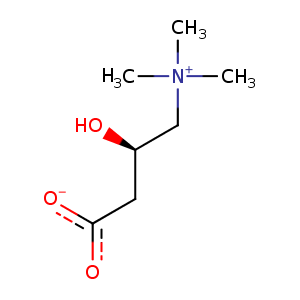
| HET Code: | 152 |
|---|---|
| Formula: | C7H15NO3 |
| Molecular weight: | 161.199 g/mol |
| DrugBank ID: | DB02648 |
| Buried Surface Area: | 60.57 % |
| Polar Surface area: | 60.36 Å2 |
| Number of | |
|---|---|
| H-Bond Acceptors: | 3 |
| H-Bond Donors: | 1 |
| Rings: | 0 |
| Aromatic rings: | 0 |
| Anionic atoms: | 1 |
| Cationic atoms: | 1 |
| Rule of Five Violation: | 0 |
| Rotatable Bonds: | 4 |
| X | Y | Z |
|---|---|---|
| 37.1657 | 15.3657 | -0.946909 |
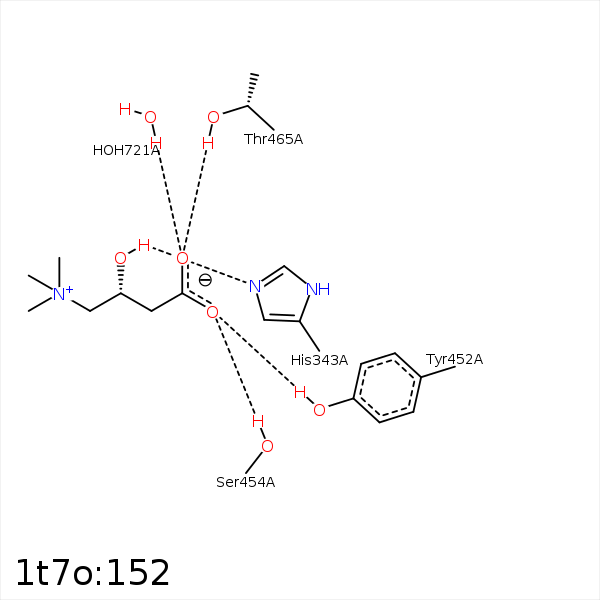


Represent the protein/ligand binding mode, centered on the ligand
Dashed lines represents hydrogen bonds and metal interactions
Green residue labels for amino acids with hydrophobic contacts (green lines) to the ligand
| Ligand | Protein | Interaction | |||
|---|---|---|---|---|---|
| Atom | Atom | Residue | Distance (Å) | Angle (°) | Type |
| O3 | NE2 | HIS- 343 | 3.24 | 165.68 | H-Bond (Ligand Donor) |
| O1B | OG | SER- 454 | 2.58 | 159.51 | H-Bond (Protein Donor) |
| C3 | CB | SER- 454 | 4.46 | 0 | Hydrophobic |
| O1A | OG1 | THR- 465 | 2.73 | 163.43 | H-Bond (Protein Donor) |
| C2 | CG2 | THR- 465 | 4.4 | 0 | Hydrophobic |
| O1A | O | HOH- 721 | 2.66 | 179.99 | H-Bond (Protein Donor) |
