sc-PDB
An Annotated Database of Druggable Binding Sites from the Protein DataBank
An Annotated Database of Druggable Binding Sites from the Protein DataBank
1.200 Å
X-ray
2004-01-07
| Name: | Aldo-keto reductase family 1 member C3 |
|---|---|
| ID: | AK1C3_HUMAN |
| AC: | P42330 |
| Organism: | Homo sapiens |
| Reign: | Eukaryota |
| TaxID: | 9606 |
| EC Number: | / |
| Chain Name: | Percentage of Residues within binding site |
|---|---|
| A | 100 % |
| B-Factor: | 8.000 |
|---|---|
| Number of residues: | 46 |
| Including | |
| Standard Amino Acids: | 44 |
| Non Standard Amino Acids: | 0 |
| Water Molecules: | 2 |
| Cofactors: | |
| Metals: | |
| Ligandability | Volume (Å3) |
|---|---|
| 0.834 | 590.625 |
| % Hydrophobic | % Polar |
|---|---|
| 50.86 | 49.14 |
| According to VolSite | |
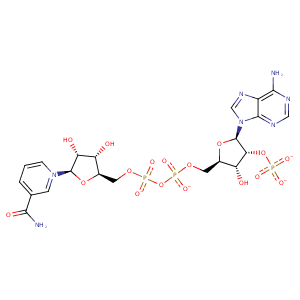
| HET Code: | NAP |
|---|---|
| Formula: | C21H25N7O17P3 |
| Molecular weight: | 740.381 g/mol |
| DrugBank ID: | DB03461 |
| Buried Surface Area: | 72.07 % |
| Polar Surface area: | 405.54 Å2 |
| Number of | |
|---|---|
| H-Bond Acceptors: | 21 |
| H-Bond Donors: | 5 |
| Rings: | 5 |
| Aromatic rings: | 3 |
| Anionic atoms: | 4 |
| Cationic atoms: | 1 |
| Rule of Five Violation: | 2 |
| Rotatable Bonds: | 13 |
| X | Y | Z |
|---|---|---|
| 28.9828 | -29.9262 | 51.8114 |


Represent the protein/ligand binding mode, centered on the ligand
Dashed lines represents hydrogen bonds and metal interactions
Green residue labels for amino acids with hydrophobic contacts (green lines) to the ligand
| Ligand | Protein | Interaction | |||
|---|---|---|---|---|---|
| Atom | Atom | Residue | Distance (Å) | Angle (°) | Type |
| O3D | N | TYR- 24 | 3.11 | 143.33 | H-Bond (Protein Donor) |
| C3D | CB | TYR- 24 | 4.11 | 0 | Hydrophobic |
| O2D | OD2 | ASP- 50 | 2.73 | 170.26 | H-Bond (Ligand Donor) |
| C2D | CZ | TYR- 55 | 3.97 | 0 | Hydrophobic |
| N7N | OG | SER- 166 | 2.81 | 138.27 | H-Bond (Ligand Donor) |
| O7N | ND2 | ASN- 167 | 2.8 | 159.61 | H-Bond (Protein Donor) |
| N7N | OE1 | GLN- 190 | 2.92 | 169.56 | H-Bond (Ligand Donor) |
| DuAr | DuAr | TYR- 216 | 3.55 | 0 | Aromatic Face/Face |
| C5N | CB | TYR- 216 | 4.07 | 0 | Hydrophobic |
| O2N | OG | SER- 217 | 2.79 | 173.24 | H-Bond (Protein Donor) |
| O5D | N | SER- 217 | 3.22 | 154.59 | H-Bond (Protein Donor) |
| O1A | N | LEU- 219 | 3.11 | 144.15 | H-Bond (Protein Donor) |
| C5B | CB | LEU- 219 | 3.95 | 0 | Hydrophobic |
| C1B | CD1 | LEU- 219 | 4.48 | 0 | Hydrophobic |
| O1A | N | SER- 221 | 3.12 | 145.25 | H-Bond (Protein Donor) |
| C5B | CG | GLN- 222 | 4.24 | 0 | Hydrophobic |
| C3B | CG | GLN- 222 | 4.19 | 0 | Hydrophobic |
| O1N | NE2 | GLN- 222 | 2.85 | 172.51 | H-Bond (Protein Donor) |
| C5D | CB | LEU- 268 | 4.4 | 0 | Hydrophobic |
| C4D | CD1 | LEU- 268 | 4.23 | 0 | Hydrophobic |
| O2A | N | LYS- 270 | 2.98 | 174.8 | H-Bond (Protein Donor) |
| O3B | NZ | LYS- 270 | 3.03 | 127.92 | H-Bond (Protein Donor) |
| O1X | NZ | LYS- 270 | 2.72 | 170.7 | H-Bond (Protein Donor) |
| C3D | CB | LYS- 270 | 4.36 | 0 | Hydrophobic |
| C3B | CD | LYS- 270 | 3.57 | 0 | Hydrophobic |
| C5D | CB | LYS- 270 | 3.76 | 0 | Hydrophobic |
| O1X | NZ | LYS- 270 | 2.72 | 0 | Ionic (Protein Cationic) |
| O3X | OG | SER- 271 | 2.65 | 169.21 | H-Bond (Protein Donor) |
| O1X | N | TYR- 272 | 2.82 | 167.66 | H-Bond (Protein Donor) |
| O2X | CZ | ARG- 276 | 3.82 | 0 | Ionic (Protein Cationic) |
| O3X | CZ | ARG- 276 | 3.55 | 0 | Ionic (Protein Cationic) |
| O2X | NH2 | ARG- 276 | 2.97 | 158 | H-Bond (Protein Donor) |
| O3X | NE | ARG- 276 | 2.71 | 175.12 | H-Bond (Protein Donor) |
| N6A | OE1 | GLN- 279 | 2.87 | 163.39 | H-Bond (Ligand Donor) |
| N7A | ND2 | ASN- 280 | 3.09 | 167.66 | H-Bond (Protein Donor) |
| N6A | OD1 | ASN- 280 | 2.84 | 150.05 | H-Bond (Ligand Donor) |
| O2A | O | HOH- 2003 | 2.74 | 179.96 | H-Bond (Protein Donor) |
