sc-PDB
An Annotated Database of Druggable Binding Sites from the Protein DataBank
An Annotated Database of Druggable Binding Sites from the Protein DataBank
2.300 Å
X-ray
2003-12-22
| Name: | Glucose--fructose oxidoreductase |
|---|---|
| ID: | GFO_ZYMMO |
| AC: | Q07982 |
| Organism: | Zymomonas mobilis subsp. mobilis |
| Reign: | Bacteria |
| TaxID: | 264203 |
| EC Number: | 1.1.99.28 |
| Chain Name: | Percentage of Residues within binding site |
|---|---|
| B | 19 % |
| C | 81 % |
| B-Factor: | 63.285 |
|---|---|
| Number of residues: | 54 |
| Including | |
| Standard Amino Acids: | 53 |
| Non Standard Amino Acids: | 0 |
| Water Molecules: | 1 |
| Cofactors: | |
| Metals: | |
| Ligandability | Volume (Å3) |
|---|---|
| 0.869 | 1147.500 |
| % Hydrophobic | % Polar |
|---|---|
| 42.65 | 57.35 |
| According to VolSite | |
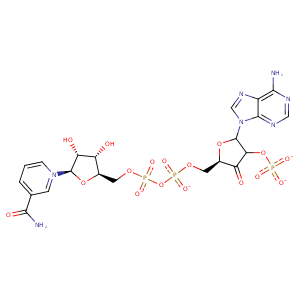
| HET Code: | NDP |
|---|---|
| Formula: | C21H26N7O17P3 |
| Molecular weight: | 741.389 g/mol |
| DrugBank ID: | DB02338 |
| Buried Surface Area: | 81.5 % |
| Polar Surface area: | 404.9 Å2 |
| Number of | |
|---|---|
| H-Bond Acceptors: | 22 |
| H-Bond Donors: | 5 |
| Rings: | 5 |
| Aromatic rings: | 2 |
| Anionic atoms: | 4 |
| Cationic atoms: | 0 |
| Rule of Five Violation: | 2 |
| Rotatable Bonds: | 13 |
| X | Y | Z |
|---|---|---|
| 28.3597 | -8.27562 | 52.5734 |


Represent the protein/ligand binding mode, centered on the ligand
Dashed lines represents hydrogen bonds and metal interactions
Green residue labels for amino acids with hydrophobic contacts (green lines) to the ligand
| Ligand | Protein | Interaction | |||
|---|---|---|---|---|---|
| Atom | Atom | Residue | Distance (Å) | Angle (°) | Type |
| N6A | O | PRO- 11 | 2.69 | 141.17 | H-Bond (Ligand Donor) |
| N6A | O | THR- 13 | 3.01 | 132.88 | H-Bond (Ligand Donor) |
| N7A | N | ALA- 15 | 3.29 | 148.95 | H-Bond (Protein Donor) |
| C2B | CB | ARG- 17 | 3.96 | 0 | Hydrophobic |
| O2B | N | LEU- 39 | 2.79 | 135.31 | H-Bond (Protein Donor) |
| O2X | N | LEU- 39 | 3.13 | 152.03 | H-Bond (Protein Donor) |
| O1A | N | LYS- 41 | 3.31 | 160.01 | H-Bond (Protein Donor) |
| C4N | CD1 | TYR- 42 | 3.38 | 0 | Hydrophobic |
| C5N | CB | TYR- 42 | 3.7 | 0 | Hydrophobic |
| O2X | OG | SER- 64 | 2.64 | 165.93 | H-Bond (Protein Donor) |
| O1X | N | GLY- 65 | 2.87 | 139.73 | H-Bond (Protein Donor) |
| O2X | NZ | LYS- 69 | 2.98 | 144.34 | H-Bond (Protein Donor) |
| O3X | NZ | LYS- 69 | 3.17 | 142.03 | H-Bond (Protein Donor) |
| O2X | NZ | LYS- 69 | 2.98 | 0 | Ionic (Protein Cationic) |
| O3X | NZ | LYS- 69 | 3.17 | 0 | Ionic (Protein Cationic) |
| O2B | OH | TYR- 87 | 3.25 | 143.92 | H-Bond (Protein Donor) |
| O1X | OH | TYR- 87 | 2.82 | 140.66 | H-Bond (Protein Donor) |
| C5D | CG2 | ILE- 105 | 3.79 | 0 | Hydrophobic |
| C4B | CD2 | LEU- 106 | 4.36 | 0 | Hydrophobic |
| C1B | CD2 | LEU- 106 | 3.5 | 0 | Hydrophobic |
| O3D | O | LEU- 106 | 3.03 | 167.65 | H-Bond (Ligand Donor) |
| O3D | NE2 | HIS- 111 | 3.13 | 121.36 | H-Bond (Protein Donor) |
| C4D | CG | GLU- 128 | 4.27 | 0 | Hydrophobic |
| N7N | OE1 | GLU- 128 | 2.79 | 156.65 | H-Bond (Ligand Donor) |
| O2D | NZ | LYS- 129 | 3 | 160.17 | H-Bond (Protein Donor) |
| O2D | O | LYS- 129 | 3.06 | 158.21 | H-Bond (Ligand Donor) |
| DuAr | NZ | LYS- 129 | 3.9 | 43.35 | Pi/Cation |
| O7N | NE | ARG- 157 | 2.6 | 137.59 | H-Bond (Protein Donor) |
| O3 | NE1 | TRP- 199 | 3.2 | 164.37 | H-Bond (Protein Donor) |
| C4D | CZ2 | TRP- 199 | 4.36 | 0 | Hydrophobic |
| C3D | CZ2 | TRP- 199 | 3.38 | 0 | Hydrophobic |
| O1N | CZ | ARG- 200 | 3.46 | 0 | Ionic (Protein Cationic) |
| O1N | NH2 | ARG- 200 | 2.75 | 166.4 | H-Bond (Protein Donor) |
| O1N | NH1 | ARG- 200 | 3.31 | 132.15 | H-Bond (Protein Donor) |
| O5D | NH2 | ARG- 200 | 3.26 | 121.1 | H-Bond (Protein Donor) |
| O2D | O | HOH- 509 | 2.72 | 179.94 | H-Bond (Protein Donor) |
