sc-PDB
An Annotated Database of Druggable Binding Sites from the Protein DataBank
An Annotated Database of Druggable Binding Sites from the Protein DataBank
1.800 Å
X-ray
1996-09-19
| Name: | Dihydrofolate reductase |
|---|---|
| ID: | DYR_ECOLI |
| AC: | P0ABQ4 |
| Organism: | Escherichia coli |
| Reign: | Bacteria |
| TaxID: | 83333 |
| EC Number: | 1.5.1.3 |
| Chain Name: | Percentage of Residues within binding site |
|---|---|
| A | 100 % |
| B-Factor: | 17.067 |
|---|---|
| Number of residues: | 35 |
| Including | |
| Standard Amino Acids: | 32 |
| Non Standard Amino Acids: | 1 |
| Water Molecules: | 2 |
| Cofactors: | NAP |
| Metals: | |
| Ligandability | Volume (Å3) |
|---|---|
| 1.290 | 334.125 |
| % Hydrophobic | % Polar |
|---|---|
| 70.71 | 29.29 |
| According to VolSite | |
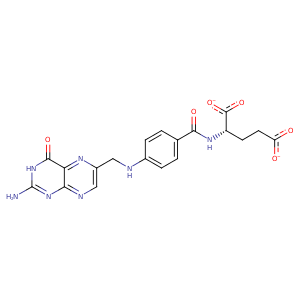
| HET Code: | FOL |
|---|---|
| Formula: | C19H17N7O6 |
| Molecular weight: | 439.382 g/mol |
| DrugBank ID: | DB00158 |
| Buried Surface Area: | 57.73 % |
| Polar Surface area: | 214.64 Å2 |
| Number of | |
|---|---|
| H-Bond Acceptors: | 12 |
| H-Bond Donors: | 4 |
| Rings: | 3 |
| Aromatic rings: | 2 |
| Anionic atoms: | 2 |
| Cationic atoms: | 0 |
| Rule of Five Violation: | 1 |
| Rotatable Bonds: | 9 |
| X | Y | Z |
|---|---|---|
| 33.0957 | 42.3424 | 7.70041 |
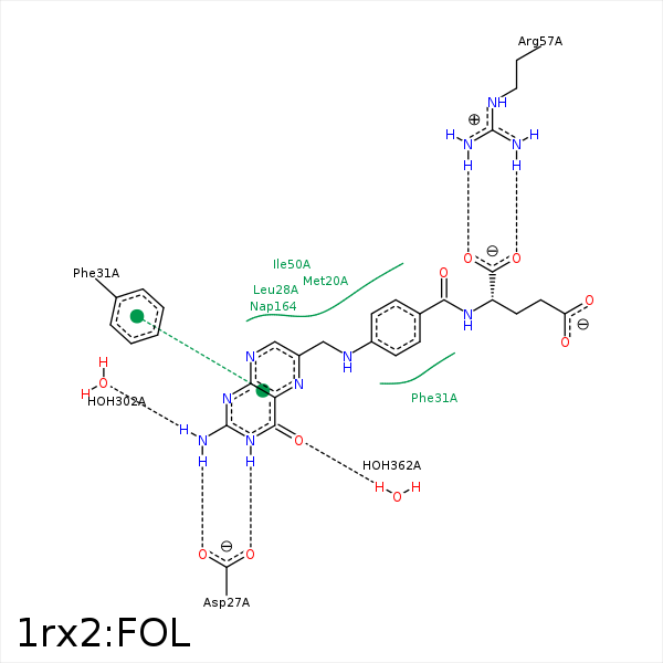


Represent the protein/ligand binding mode, centered on the ligand
Dashed lines represents hydrogen bonds and metal interactions
Green residue labels for amino acids with hydrophobic contacts (green lines) to the ligand
| Ligand | Protein | Interaction | |||
|---|---|---|---|---|---|
| Atom | Atom | Residue | Distance (Å) | Angle (°) | Type |
| C13 | CE | MET- 20 | 4.06 | 0 | Hydrophobic |
| NA2 | OD1 | ASP- 27 | 2.97 | 169.34 | H-Bond (Ligand Donor) |
| N3 | OD2 | ASP- 27 | 2.62 | 168.65 | H-Bond (Ligand Donor) |
| CB | CB | LEU- 28 | 4.39 | 0 | Hydrophobic |
| C11 | CD2 | LEU- 28 | 4.26 | 0 | Hydrophobic |
| C16 | CE2 | PHE- 31 | 3.43 | 0 | Hydrophobic |
| CB | CB | LYS- 32 | 4.21 | 0 | Hydrophobic |
| C9 | CG2 | THR- 46 | 4.1 | 0 | Hydrophobic |
| C9 | CD1 | ILE- 50 | 4.09 | 0 | Hydrophobic |
| C15 | CD1 | ILE- 50 | 4 | 0 | Hydrophobic |
| C14 | CG1 | ILE- 50 | 3.88 | 0 | Hydrophobic |
| C16 | CD2 | LEU- 54 | 4.23 | 0 | Hydrophobic |
| O1 | CZ | ARG- 57 | 3.48 | 0 | Ionic (Protein Cationic) |
| O2 | CZ | ARG- 57 | 3.51 | 0 | Ionic (Protein Cationic) |
| O1 | NH2 | ARG- 57 | 3.39 | 128.44 | H-Bond (Protein Donor) |
| O1 | NH1 | ARG- 57 | 2.7 | 163.35 | H-Bond (Protein Donor) |
| O2 | NH2 | ARG- 57 | 2.63 | 174.16 | H-Bond (Protein Donor) |
| C9 | C4N | NAP- 164 | 3.67 | 0 | Hydrophobic |
| NA2 | O | HOH- 302 | 2.93 | 140.63 | H-Bond (Ligand Donor) |
| O4 | O | HOH- 362 | 2.82 | 179.96 | H-Bond (Protein Donor) |
