sc-PDB
An Annotated Database of Druggable Binding Sites from the Protein DataBank
An Annotated Database of Druggable Binding Sites from the Protein DataBank
2.800 Å
X-ray
2003-11-04
| Name: | Purine nucleoside phosphorylase |
|---|---|
| ID: | PNPH_HUMAN |
| AC: | P00491 |
| Organism: | Homo sapiens |
| Reign: | Eukaryota |
| TaxID: | 9606 |
| EC Number: | 2.4.2.1 |
| Chain Name: | Percentage of Residues within binding site |
|---|---|
| E | 100 % |
| B-Factor: | 41.484 |
|---|---|
| Number of residues: | 29 |
| Including | |
| Standard Amino Acids: | 29 |
| Non Standard Amino Acids: | 0 |
| Water Molecules: | 0 |
| Cofactors: | |
| Metals: | |
| Ligandability | Volume (Å3) |
|---|---|
| 0.810 | 324.000 |
| % Hydrophobic | % Polar |
|---|---|
| 40.63 | 59.38 |
| According to VolSite | |
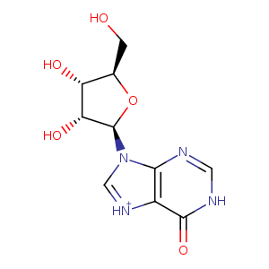
| HET Code: | NOS |
|---|---|
| Formula: | C10H12N4O5 |
| Molecular weight: | 268.226 g/mol |
| DrugBank ID: | DB04335 |
| Buried Surface Area: | 71.19 % |
| Polar Surface area: | 133.75 Å2 |
| Number of | |
|---|---|
| H-Bond Acceptors: | 8 |
| H-Bond Donors: | 4 |
| Rings: | 3 |
| Aromatic rings: | 2 |
| Anionic atoms: | 0 |
| Cationic atoms: | 0 |
| Rule of Five Violation: | 0 |
| Rotatable Bonds: | 2 |
| X | Y | Z |
|---|---|---|
| 53.6515 | 129.301 | 55.1294 |
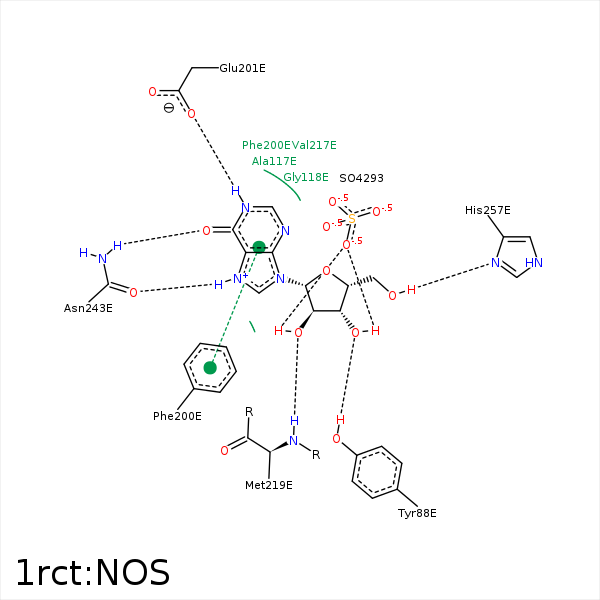


Represent the protein/ligand binding mode, centered on the ligand
Dashed lines represents hydrogen bonds and metal interactions
Green residue labels for amino acids with hydrophobic contacts (green lines) to the ligand
| Ligand | Protein | Interaction | |||
|---|---|---|---|---|---|
| Atom | Atom | Residue | Distance (Å) | Angle (°) | Type |
| O3' | OH | TYR- 88 | 3.03 | 164.41 | H-Bond (Protein Donor) |
| C1' | CB | ALA- 116 | 4.29 | 0 | Hydrophobic |
| N1 | OE1 | GLU- 201 | 3.15 | 121.52 | H-Bond (Ligand Donor) |
| N1 | OE2 | GLU- 201 | 2.5 | 161.47 | H-Bond (Ligand Donor) |
| C2' | CG | MET- 219 | 3.88 | 0 | Hydrophobic |
| C3' | SD | MET- 219 | 3.93 | 0 | Hydrophobic |
| O2' | N | MET- 219 | 3.05 | 142.31 | H-Bond (Protein Donor) |
| O6 | ND2 | ASN- 243 | 2.87 | 156.92 | H-Bond (Protein Donor) |
| C5' | CB | HIS- 257 | 3.39 | 0 | Hydrophobic |
| O5' | ND1 | HIS- 257 | 2.99 | 174.99 | H-Bond (Ligand Donor) |
