sc-PDB
An Annotated Database of Druggable Binding Sites from the Protein DataBank
An Annotated Database of Druggable Binding Sites from the Protein DataBank
2.400 Å
X-ray
2003-02-19
| Name: | Purine nucleoside phosphorylase |
|---|---|
| ID: | Q5SID9_THET8 |
| AC: | Q5SID9 |
| Organism: | Thermus thermophilus |
| Reign: | Bacteria |
| TaxID: | 300852 |
| EC Number: | / |
| Chain Name: | Percentage of Residues within binding site |
|---|---|
| C | 88 % |
| D | 12 % |
| B-Factor: | 29.875 |
|---|---|
| Number of residues: | 33 |
| Including | |
| Standard Amino Acids: | 32 |
| Non Standard Amino Acids: | 0 |
| Water Molecules: | 1 |
| Cofactors: | |
| Metals: | |
| Ligandability | Volume (Å3) |
|---|---|
| 0.643 | 958.500 |
| % Hydrophobic | % Polar |
|---|---|
| 37.68 | 62.32 |
| According to VolSite | |
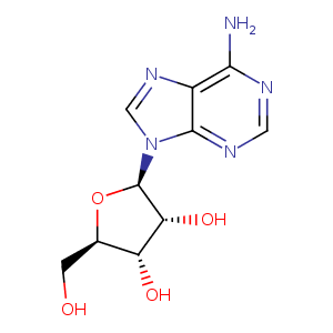
| HET Code: | ADN |
|---|---|
| Formula: | C10H13N5O4 |
| Molecular weight: | 267.241 g/mol |
| DrugBank ID: | DB00640 |
| Buried Surface Area: | 75.09 % |
| Polar Surface area: | 139.54 Å2 |
| Number of | |
|---|---|
| H-Bond Acceptors: | 8 |
| H-Bond Donors: | 4 |
| Rings: | 3 |
| Aromatic rings: | 2 |
| Anionic atoms: | 0 |
| Cationic atoms: | 0 |
| Rule of Five Violation: | 0 |
| Rotatable Bonds: | 2 |
| X | Y | Z |
|---|---|---|
| 7.06126 | -122.953 | -139.361 |
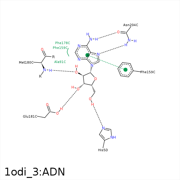


Represent the protein/ligand binding mode, centered on the ligand
Dashed lines represents hydrogen bonds and metal interactions
Green residue labels for amino acids with hydrophobic contacts (green lines) to the ligand
| Ligand | Protein | Interaction | |||
|---|---|---|---|---|---|
| Atom | Atom | Residue | Distance (Å) | Angle (°) | Type |
| C4' | SD | MET- 65 | 3.96 | 0 | Hydrophobic |
| C5' | CE1 | PHE- 159 | 3.61 | 0 | Hydrophobic |
| C2' | CB | GLU- 179 | 4.15 | 0 | Hydrophobic |
| C2' | CG | MET- 180 | 4.01 | 0 | Hydrophobic |
| C3' | SD | MET- 180 | 3.52 | 0 | Hydrophobic |
| O2' | N | MET- 180 | 3.08 | 152.16 | H-Bond (Protein Donor) |
| O3' | OE1 | GLU- 181 | 2.52 | 172.25 | H-Bond (Protein Donor) |
| N7 | ND2 | ASN- 204 | 3.1 | 163.85 | H-Bond (Protein Donor) |
| N6 | OD1 | ASN- 204 | 3.28 | 156.97 | H-Bond (Ligand Donor) |
