sc-PDB
An Annotated Database of Druggable Binding Sites from the Protein DataBank
An Annotated Database of Druggable Binding Sites from the Protein DataBank
2.450 Å
X-ray
2003-02-20
| Name: | Aldehyde dehydrogenase, mitochondrial |
|---|---|
| ID: | ALDH2_HUMAN |
| AC: | P05091 |
| Organism: | Homo sapiens |
| Reign: | Eukaryota |
| TaxID: | 9606 |
| EC Number: | 1.2.1.3 |
| Chain Name: | Percentage of Residues within binding site |
|---|---|
| B | 100 % |
| B-Factor: | 39.298 |
|---|---|
| Number of residues: | 52 |
| Including | |
| Standard Amino Acids: | 49 |
| Non Standard Amino Acids: | 1 |
| Water Molecules: | 2 |
| Cofactors: | |
| Metals: | MG |
| Ligandability | Volume (Å3) |
|---|---|
| 0.414 | 810.000 |
| % Hydrophobic | % Polar |
|---|---|
| 44.17 | 55.83 |
| According to VolSite | |
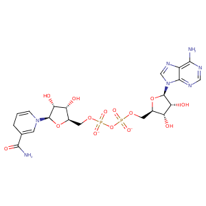
| HET Code: | NAI |
|---|---|
| Formula: | C21H27N7O14P2 |
| Molecular weight: | 663.425 g/mol |
| DrugBank ID: | DB00157 |
| Buried Surface Area: | 71.57 % |
| Polar Surface area: | 342.9 Å2 |
| Number of | |
|---|---|
| H-Bond Acceptors: | 19 |
| H-Bond Donors: | 6 |
| Rings: | 5 |
| Aromatic rings: | 2 |
| Anionic atoms: | 2 |
| Cationic atoms: | 0 |
| Rule of Five Violation: | 3 |
| Rotatable Bonds: | 11 |
| X | Y | Z |
|---|---|---|
| 64.5493 | 46.0829 | 31.1259 |
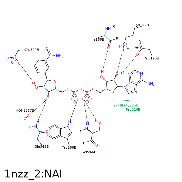


Represent the protein/ligand binding mode, centered on the ligand
Dashed lines represents hydrogen bonds and metal interactions
Green residue labels for amino acids with hydrophobic contacts (green lines) to the ligand
| Ligand | Protein | Interaction | |||
|---|---|---|---|---|---|
| Atom | Atom | Residue | Distance (Å) | Angle (°) | Type |
| C1B | CG2 | ILE- 165 | 3.75 | 0 | Hydrophobic |
| C4B | CG2 | ILE- 165 | 4.15 | 0 | Hydrophobic |
| O3B | O | ILE- 166 | 3.08 | 150.18 | H-Bond (Ligand Donor) |
| C4N | CB | PRO- 167 | 3.47 | 0 | Hydrophobic |
| O2N | NE1 | TRP- 168 | 3.03 | 162.89 | H-Bond (Protein Donor) |
| O7N | ND2 | ASN- 169 | 3.38 | 136.47 | H-Bond (Protein Donor) |
| O2B | NZ | LYS- 192 | 2.79 | 158.93 | H-Bond (Protein Donor) |
| C3B | CB | ALA- 194 | 4.42 | 0 | Hydrophobic |
| O2B | OE1 | GLU- 195 | 3.05 | 146.97 | H-Bond (Ligand Donor) |
| C4B | CE1 | PHE- 243 | 4.03 | 0 | Hydrophobic |
| C1B | CE1 | PHE- 243 | 4.17 | 0 | Hydrophobic |
| N7N | O | GLY- 245 | 3.31 | 122.95 | H-Bond (Ligand Donor) |
| O1A | N | SER- 246 | 3.22 | 168.66 | H-Bond (Protein Donor) |
| O1A | OG | SER- 246 | 3.15 | 165.55 | H-Bond (Protein Donor) |
| C1D | CB | SER- 246 | 4.02 | 0 | Hydrophobic |
| C4D | CB | SER- 246 | 3.85 | 0 | Hydrophobic |
| O3D | NE2 | GLN- 349 | 3.1 | 130.86 | H-Bond (Protein Donor) |
| O2D | OE1 | GLU- 399 | 2.64 | 159.52 | H-Bond (Ligand Donor) |
| C3D | CD2 | PHE- 401 | 3.97 | 0 | Hydrophobic |
| C2D | CG | PHE- 401 | 3.69 | 0 | Hydrophobic |
| C4N | CZ | PHE- 401 | 3.63 | 0 | Hydrophobic |
| O2A | MG | MG- 1602 | 2.08 | 0 | Metal Acceptor |
| O1N | MG | MG- 1602 | 2.24 | 0 | Metal Acceptor |
| O3D | O | HOH- 2567 | 3.1 | 142.52 | H-Bond (Protein Donor) |
