sc-PDB
An Annotated Database of Druggable Binding Sites from the Protein DataBank
An Annotated Database of Druggable Binding Sites from the Protein DataBank
2.500 Å
X-ray
2002-07-12
| Name: | Glycerol-3-phosphate dehydrogenase [NAD(+)], glycosomal |
|---|---|
| ID: | GPDA_LEIME |
| AC: | P90551 |
| Organism: | Leishmania mexicana |
| Reign: | Eukaryota |
| TaxID: | 5665 |
| EC Number: | 1.1.1.8 |
| Chain Name: | Percentage of Residues within binding site |
|---|---|
| A | 100 % |
| B-Factor: | 47.745 |
|---|---|
| Number of residues: | 8 |
| Including | |
| Standard Amino Acids: | 8 |
| Non Standard Amino Acids: | 0 |
| Water Molecules: | 0 |
| Cofactors: | |
| Metals: | |
| Ligandability | Volume (Å3) |
|---|---|
| 1.048 | 313.875 |
| % Hydrophobic | % Polar |
|---|---|
| 68.82 | 31.18 |
| According to VolSite | |
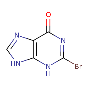
| HET Code: | BOA |
|---|---|
| Formula: | C5H3BrN4O |
| Molecular weight: | 215.008 g/mol |
| DrugBank ID: | DB04283 |
| Buried Surface Area: | 34.66 % |
| Polar Surface area: | 74.69 Å2 |
| Number of | |
|---|---|
| H-Bond Acceptors: | 4 |
| H-Bond Donors: | 2 |
| Rings: | 2 |
| Aromatic rings: | 2 |
| Anionic atoms: | 0 |
| Cationic atoms: | 0 |
| Rule of Five Violation: | 0 |
| Rotatable Bonds: | 0 |
| X | Y | Z |
|---|---|---|
| 34.2224 | 54.7654 | 30.1223 |
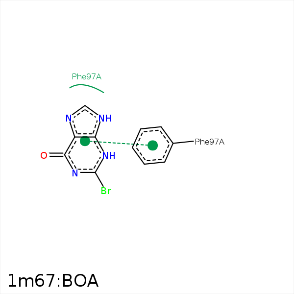


Represent the protein/ligand binding mode, centered on the ligand
Dashed lines represents hydrogen bonds and metal interactions
Green residue labels for amino acids with hydrophobic contacts (green lines) to the ligand
| Ligand | Protein | Interaction | |||
|---|---|---|---|---|---|
| Atom | Atom | Residue | Distance (Å) | Angle (°) | Type |
| BR | CZ3 | TRP- 44 | 3.93 | 0 | Hydrophobic |
| BR | CG2 | ILE- 93 | 3.89 | 0 | Hydrophobic |
| BR | CB | PHE- 97 | 3.89 | 0 | Hydrophobic |
| DuAr | DuAr | PHE- 97 | 3.54 | 0 | Aromatic Face/Face |
| BR | CB | PHE- 101 | 4.27 | 0 | Hydrophobic |
