sc-PDB
An Annotated Database of Druggable Binding Sites from the Protein DataBank
An Annotated Database of Druggable Binding Sites from the Protein DataBank
2.300 Å
X-ray
2000-12-27
| Name: | UDP-N-acetylenolpyruvoylglucosamine reductase |
|---|---|
| ID: | MURB_STAAU |
| AC: | P61431 |
| Organism: | Staphylococcus aureus |
| Reign: | Bacteria |
| TaxID: | 1280 |
| EC Number: | / |
| Chain Name: | Percentage of Residues within binding site |
|---|---|
| A | 100 % |
| B-Factor: | 26.375 |
|---|---|
| Number of residues: | 59 |
| Including | |
| Standard Amino Acids: | 54 |
| Non Standard Amino Acids: | 0 |
| Water Molecules: | 5 |
| Cofactors: | |
| Metals: | |
| Ligandability | Volume (Å3) |
|---|---|
| 1.102 | 1302.750 |
| % Hydrophobic | % Polar |
|---|---|
| 41.45 | 58.55 |
| According to VolSite | |
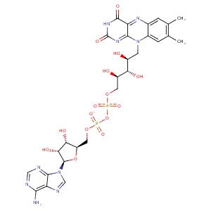
| HET Code: | FAD |
|---|---|
| Formula: | C27H31N9O15P2 |
| Molecular weight: | 783.534 g/mol |
| DrugBank ID: | DB03147 |
| Buried Surface Area: | 78.27 % |
| Polar Surface area: | 381.7 Å2 |
| Number of | |
|---|---|
| H-Bond Acceptors: | 22 |
| H-Bond Donors: | 7 |
| Rings: | 6 |
| Aromatic rings: | 3 |
| Anionic atoms: | 2 |
| Cationic atoms: | 0 |
| Rule of Five Violation: | 3 |
| Rotatable Bonds: | 13 |
| X | Y | Z |
|---|---|---|
| 179.772 | 149.015 | 163.653 |
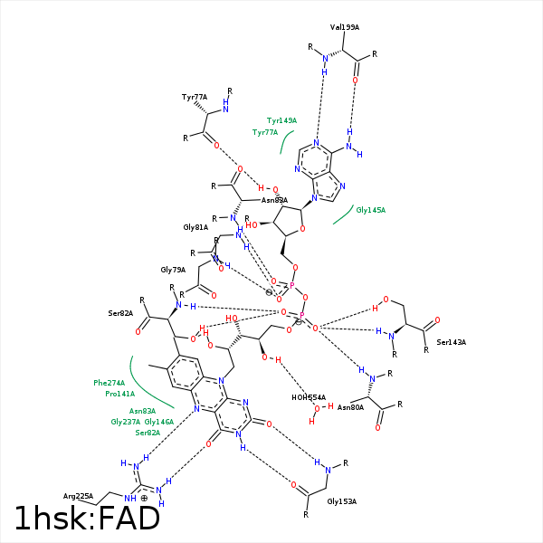


Represent the protein/ligand binding mode, centered on the ligand
Dashed lines represents hydrogen bonds and metal interactions
Green residue labels for amino acids with hydrophobic contacts (green lines) to the ligand
| Ligand | Protein | Interaction | |||
|---|---|---|---|---|---|
| Atom | Atom | Residue | Distance (Å) | Angle (°) | Type |
| C8M | CE2 | TYR- 42 | 3.3 | 0 | Hydrophobic |
| O2B | O | TYR- 77 | 2.68 | 154.76 | H-Bond (Ligand Donor) |
| O1A | N | GLY- 79 | 2.86 | 142.53 | H-Bond (Protein Donor) |
| O1P | N | ASN- 80 | 2.98 | 169.42 | H-Bond (Protein Donor) |
| O1A | N | GLY- 81 | 2.72 | 144.17 | H-Bond (Protein Donor) |
| C2' | CB | SER- 82 | 4.29 | 0 | Hydrophobic |
| O2' | OG | SER- 82 | 2.93 | 151.05 | H-Bond (Ligand Donor) |
| O2P | N | SER- 82 | 2.83 | 156.71 | H-Bond (Protein Donor) |
| O2A | N | ASN- 83 | 2.73 | 153.71 | H-Bond (Protein Donor) |
| C5B | CB | ASN- 83 | 4.19 | 0 | Hydrophobic |
| C5' | CB | ASN- 83 | 4.06 | 0 | Hydrophobic |
| C2' | CB | ASN- 83 | 3.91 | 0 | Hydrophobic |
| C5B | CD1 | ILE- 84 | 4.08 | 0 | Hydrophobic |
| C3B | CG2 | ILE- 84 | 3.92 | 0 | Hydrophobic |
| C7 | CG | PRO- 141 | 3.73 | 0 | Hydrophobic |
| C8 | CG | PRO- 141 | 3.79 | 0 | Hydrophobic |
| C8 | CG | PRO- 141 | 3.79 | 0 | Hydrophobic |
| O1P | N | SER- 143 | 2.92 | 168.72 | H-Bond (Protein Donor) |
| O1P | OG | SER- 143 | 2.69 | 165.27 | H-Bond (Protein Donor) |
| C4B | CE2 | TYR- 149 | 3.79 | 0 | Hydrophobic |
| C3B | CZ | TYR- 149 | 4.31 | 0 | Hydrophobic |
| C1B | CD1 | TYR- 149 | 3.72 | 0 | Hydrophobic |
| O3B | OH | TYR- 149 | 3.26 | 120.74 | H-Bond (Ligand Donor) |
| O2 | N | GLY- 153 | 2.88 | 150.86 | H-Bond (Protein Donor) |
| N3 | O | GLY- 153 | 2.65 | 169.08 | H-Bond (Ligand Donor) |
| N6A | O | VAL- 199 | 3.05 | 161.69 | H-Bond (Ligand Donor) |
| N1A | N | VAL- 199 | 2.79 | 170.95 | H-Bond (Protein Donor) |
| O4 | NH1 | ARG- 225 | 3.07 | 166.53 | H-Bond (Protein Donor) |
| N5 | NH2 | ARG- 225 | 3.23 | 141.98 | H-Bond (Protein Donor) |
| C7M | CD2 | LEU- 231 | 3.92 | 0 | Hydrophobic |
| C7M | CE2 | PHE- 274 | 3.33 | 0 | Hydrophobic |
| O4' | O | HOH- 554 | 2.88 | 163.34 | H-Bond (Ligand Donor) |
