sc-PDB
An Annotated Database of Druggable Binding Sites from the Protein DataBank
An Annotated Database of Druggable Binding Sites from the Protein DataBank
2.000 Å
X-ray
2001-05-22
| Name: | Dimethyl sulfoxide/trimethylamine N-oxide reductase |
|---|---|
| ID: | DSTOR_RHOCA |
| AC: | Q52675 |
| Organism: | Rhodobacter capsulatus |
| Reign: | Bacteria |
| TaxID: | 1061 |
| EC Number: | 1.7.2.3 |
| Chain Name: | Percentage of Residues within binding site |
|---|---|
| C | 100 % |
| B-Factor: | 12.943 |
|---|---|
| Number of residues: | 64 |
| Including | |
| Standard Amino Acids: | 58 |
| Non Standard Amino Acids: | 1 |
| Water Molecules: | 5 |
| Cofactors: | |
| Metals: | |
| Ligandability | Volume (Å3) |
|---|---|
| 0.805 | 526.500 |
| % Hydrophobic | % Polar |
|---|---|
| 50.00 | 50.00 |
| According to VolSite | |
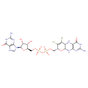
| HET Code: | PGD |
|---|---|
| Formula: | C20H24N10O13P2S2 |
| Molecular weight: | 738.541 g/mol |
| DrugBank ID: | - |
| Buried Surface Area: | 82.43 % |
| Polar Surface area: | 444.81 Å2 |
| Number of | |
|---|---|
| H-Bond Acceptors: | 19 |
| H-Bond Donors: | 10 |
| Rings: | 6 |
| Aromatic rings: | 1 |
| Anionic atoms: | 2 |
| Cationic atoms: | 2 |
| Rule of Five Violation: | 3 |
| Rotatable Bonds: | 9 |
| X | Y | Z |
|---|---|---|
| 8.67223 | 64.565 | -23.835 |


Represent the protein/ligand binding mode, centered on the ligand
Dashed lines represents hydrogen bonds and metal interactions
Green residue labels for amino acids with hydrophobic contacts (green lines) to the ligand
| Ligand | Protein | Interaction | |||
|---|---|---|---|---|---|
| Atom | Atom | Residue | Distance (Å) | Angle (°) | Type |
| S12 | CE2 | TYR- 114 | 3.84 | 0 | Hydrophobic |
| C11 | CE2 | TYR- 114 | 4.03 | 0 | Hydrophobic |
| O2A | N | TRP- 116 | 2.64 | 138.91 | H-Bond (Protein Donor) |
| C11 | CB | TYR- 146 | 4.02 | 0 | Hydrophobic |
| S12 | CB | TYR- 146 | 4.32 | 0 | Hydrophobic |
| S13 | CB | TYR- 146 | 4.01 | 0 | Hydrophobic |
| C14 | CB | TYR- 146 | 3.61 | 0 | Hydrophobic |
| C23 | CD2 | TYR- 146 | 3.73 | 0 | Hydrophobic |
| S13 | CB | SER- 147 | 3.83 | 0 | Hydrophobic |
| S13 | CD | ARG- 326 | 4.39 | 0 | Hydrophobic |
| C14 | CD | ARG- 326 | 4.27 | 0 | Hydrophobic |
| O17 | NH1 | ARG- 326 | 3.36 | 130.36 | H-Bond (Protein Donor) |
| N3 | N | GLY- 432 | 3.47 | 124.47 | H-Bond (Protein Donor) |
| O1A | N | ASN- 434 | 2.58 | 157.12 | H-Bond (Protein Donor) |
| N22 | O | HIS- 438 | 2.96 | 151.74 | H-Bond (Ligand Donor) |
| C23 | CB | HIS- 438 | 3.85 | 0 | Hydrophobic |
| C11 | CB | HIS- 438 | 3.86 | 0 | Hydrophobic |
| N20 | NE2 | GLN- 440 | 3.35 | 121.49 | H-Bond (Protein Donor) |
| N2 | O | HIS- 458 | 3.07 | 178.93 | H-Bond (Ligand Donor) |
| O3' | OD2 | ASP- 459 | 2.69 | 162.73 | H-Bond (Ligand Donor) |
| O2' | OD1 | ASP- 459 | 2.74 | 169.33 | H-Bond (Ligand Donor) |
| O6 | NH1 | ARG- 481 | 2.91 | 166 | H-Bond (Protein Donor) |
| N1 | OD2 | ASP- 511 | 2.73 | 172.97 | H-Bond (Ligand Donor) |
| N2 | OD2 | ASP- 511 | 3.5 | 127.92 | H-Bond (Ligand Donor) |
| N2 | OD1 | ASP- 511 | 2.77 | 162.46 | H-Bond (Ligand Donor) |
| N18 | O | ALA- 641 | 2.98 | 134.94 | H-Bond (Ligand Donor) |
| N15 | NE2 | HIS- 643 | 3.03 | 156.11 | H-Bond (Ligand Donor) |
| C10 | CB | HIS- 649 | 4.21 | 0 | Hydrophobic |
| O1B | N | SER- 650 | 2.71 | 139.44 | H-Bond (Protein Donor) |
| O2B | NE2 | GLN- 651 | 2.75 | 164.38 | H-Bond (Protein Donor) |
| C5' | CG | GLN- 651 | 4.11 | 0 | Hydrophobic |
| C3' | CG | GLN- 651 | 3.81 | 0 | Hydrophobic |
| N19 | OD1 | ASN- 737 | 2.92 | 165.41 | H-Bond (Ligand Donor) |
| N20 | ND2 | ASN- 737 | 3.11 | 160.3 | H-Bond (Protein Donor) |
| O17 | NE2 | GLN- 755 | 3.19 | 155.96 | H-Bond (Protein Donor) |
| O6 | O | HOH- 2577 | 2.79 | 179.99 | H-Bond (Protein Donor) |
