sc-PDB
An Annotated Database of Druggable Binding Sites from the Protein DataBank
An Annotated Database of Druggable Binding Sites from the Protein DataBank
2.500 Å
X-ray
1997-03-22
| Name: | Fibroblast growth factor receptor 1 |
|---|---|
| ID: | FGFR1_HUMAN |
| AC: | P11362 |
| Organism: | Homo sapiens |
| Reign: | Eukaryota |
| TaxID: | 9606 |
| EC Number: | / |
| Chain Name: | Percentage of Residues within binding site |
|---|---|
| A | 100 % |
| B-Factor: | 34.196 |
|---|---|
| Number of residues: | 29 |
| Including | |
| Standard Amino Acids: | 29 |
| Non Standard Amino Acids: | 0 |
| Water Molecules: | 0 |
| Cofactors: | |
| Metals: | |
| Ligandability | Volume (Å3) |
|---|---|
| 1.085 | 678.375 |
| % Hydrophobic | % Polar |
|---|---|
| 60.70 | 39.30 |
| According to VolSite | |
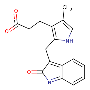
| HET Code: | SU1 |
|---|---|
| Formula: | C17H15N2O3 |
| Molecular weight: | 295.313 g/mol |
| DrugBank ID: | DB08577 |
| Buried Surface Area: | 65.27 % |
| Polar Surface area: | 85.02 Å2 |
| Number of | |
|---|---|
| H-Bond Acceptors: | 3 |
| H-Bond Donors: | 2 |
| Rings: | 3 |
| Aromatic rings: | 2 |
| Anionic atoms: | 1 |
| Cationic atoms: | 0 |
| Rule of Five Violation: | 0 |
| Rotatable Bonds: | 4 |
| X | Y | Z |
|---|---|---|
| 8.71095 | 4.01168 | 21.7269 |
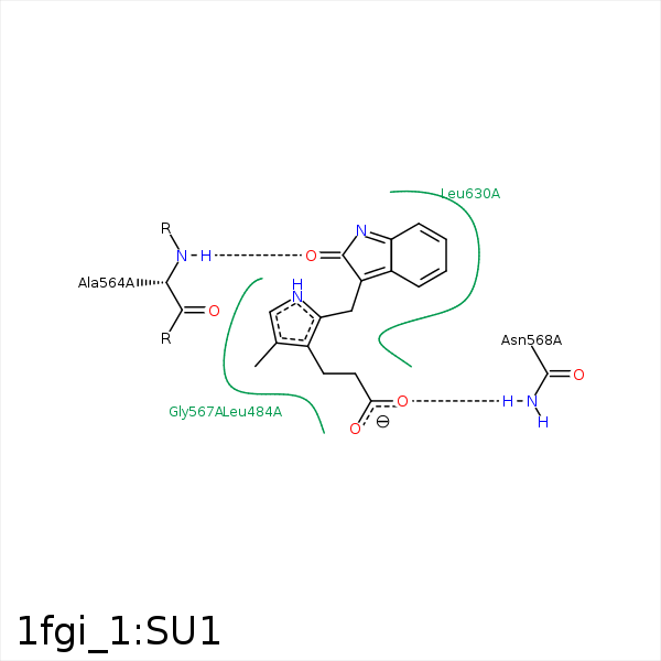


Represent the protein/ligand binding mode, centered on the ligand
Dashed lines represents hydrogen bonds and metal interactions
Green residue labels for amino acids with hydrophobic contacts (green lines) to the ligand
| Ligand | Protein | Interaction | |||
|---|---|---|---|---|---|
| Atom | Atom | Residue | Distance (Å) | Angle (°) | Type |
| C10 | CB | LEU- 484 | 4.06 | 0 | Hydrophobic |
| C12 | CB | LEU- 484 | 4.39 | 0 | Hydrophobic |
| C13 | CE2 | PHE- 489 | 4.32 | 0 | Hydrophobic |
| O2 | N | ALA- 564 | 2.76 | 171.11 | H-Bond (Protein Donor) |
| O4 | ND2 | ASN- 568 | 2.78 | 165.58 | H-Bond (Protein Donor) |
| C10 | CD1 | LEU- 630 | 4.34 | 0 | Hydrophobic |
| C13 | CD2 | LEU- 630 | 4 | 0 | Hydrophobic |
