sc-PDB
An Annotated Database of Druggable Binding Sites from the Protein DataBank
An Annotated Database of Druggable Binding Sites from the Protein DataBank
1.800 Å
X-ray
2000-03-06
| Name: | UDP-glucose 4-epimerase |
|---|---|
| ID: | GALE_HUMAN |
| AC: | Q14376 |
| Organism: | Homo sapiens |
| Reign: | Eukaryota |
| TaxID: | 9606 |
| EC Number: | / |
| Chain Name: | Percentage of Residues within binding site |
|---|---|
| A | 100 % |
| B-Factor: | 22.889 |
|---|---|
| Number of residues: | 47 |
| Including | |
| Standard Amino Acids: | 44 |
| Non Standard Amino Acids: | 0 |
| Water Molecules: | 3 |
| Cofactors: | |
| Metals: | |
| Ligandability | Volume (Å3) |
|---|---|
| 0.997 | 1451.250 |
| % Hydrophobic | % Polar |
|---|---|
| 36.51 | 63.49 |
| According to VolSite | |
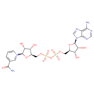
| HET Code: | NAD |
|---|---|
| Formula: | C21H26N7O14P2 |
| Molecular weight: | 662.417 g/mol |
| DrugBank ID: | - |
| Buried Surface Area: | 75.7 % |
| Polar Surface area: | 343.54 Å2 |
| Number of | |
|---|---|
| H-Bond Acceptors: | 18 |
| H-Bond Donors: | 6 |
| Rings: | 5 |
| Aromatic rings: | 3 |
| Anionic atoms: | 2 |
| Cationic atoms: | 1 |
| Rule of Five Violation: | 3 |
| Rotatable Bonds: | 11 |
| X | Y | Z |
|---|---|---|
| -2.53539 | 13.9777 | 55.1453 |
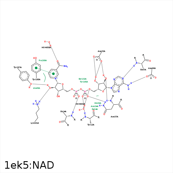


Represent the protein/ligand binding mode, centered on the ligand
Dashed lines represents hydrogen bonds and metal interactions
Green residue labels for amino acids with hydrophobic contacts (green lines) to the ligand
| Ligand | Protein | Interaction | |||
|---|---|---|---|---|---|
| Atom | Atom | Residue | Distance (Å) | Angle (°) | Type |
| O2A | N | TYR- 13 | 2.88 | 176.05 | H-Bond (Protein Donor) |
| O2N | N | ILE- 14 | 2.74 | 178.1 | H-Bond (Protein Donor) |
| C3N | CD1 | ILE- 14 | 4.28 | 0 | Hydrophobic |
| O3B | OD1 | ASP- 33 | 2.5 | 153.99 | H-Bond (Ligand Donor) |
| O3B | OD2 | ASP- 33 | 3.09 | 128.11 | H-Bond (Ligand Donor) |
| O2B | OD2 | ASP- 33 | 2.68 | 162.89 | H-Bond (Ligand Donor) |
| N3A | N | ASN- 34 | 3.31 | 138.56 | H-Bond (Protein Donor) |
| C2B | CB | HIS- 36 | 4.35 | 0 | Hydrophobic |
| O1A | ND2 | ASN- 37 | 2.93 | 167.12 | H-Bond (Protein Donor) |
| O2B | N | ASN- 37 | 2.7 | 152.77 | H-Bond (Protein Donor) |
| C2B | CB | ASN- 37 | 3.86 | 0 | Hydrophobic |
| N6A | OD1 | ASP- 66 | 2.73 | 150.55 | H-Bond (Ligand Donor) |
| N1A | N | ILE- 67 | 3.28 | 165.65 | H-Bond (Protein Donor) |
| C4D | CB | PHE- 88 | 4.17 | 0 | Hydrophobic |
| O1A | NZ | LYS- 92 | 3.81 | 0 | Ionic (Protein Cationic) |
| O3 | NZ | LYS- 92 | 3.49 | 121.94 | H-Bond (Protein Donor) |
| C5D | CG | LYS- 92 | 4.41 | 0 | Hydrophobic |
| C2D | CB | LYS- 92 | 3.69 | 0 | Hydrophobic |
| O3D | O | SER- 130 | 3.47 | 148.46 | H-Bond (Ligand Donor) |
| C5N | CB | SER- 132 | 3.94 | 0 | Hydrophobic |
| C2D | CZ | TYR- 157 | 4.34 | 0 | Hydrophobic |
| O2D | NZ | LYS- 161 | 2.75 | 169.65 | H-Bond (Protein Donor) |
| C5N | CE2 | TYR- 185 | 3.29 | 0 | Hydrophobic |
| C3N | CG | PRO- 188 | 4.2 | 0 | Hydrophobic |
| O5B | O | HOH- 601 | 3.13 | 153.2 | H-Bond (Protein Donor) |
| O7N | O | HOH- 658 | 2.74 | 179.96 | H-Bond (Protein Donor) |
