sc-PDB
An Annotated Database of Druggable Binding Sites from the Protein DataBank
An Annotated Database of Druggable Binding Sites from the Protein DataBank
2.300 Å
X-ray
2000-02-23
| Name: | DNA gyrase subunit B |
|---|---|
| ID: | GYRB_ECOLI |
| AC: | P0AES6 |
| Organism: | Escherichia coli |
| Reign: | Bacteria |
| TaxID: | 83333 |
| EC Number: | / |
| Chain Name: | Percentage of Residues within binding site |
|---|---|
| A | 95 % |
| B | 5 % |
| B-Factor: | 19.014 |
|---|---|
| Number of residues: | 47 |
| Including | |
| Standard Amino Acids: | 44 |
| Non Standard Amino Acids: | 0 |
| Water Molecules: | 3 |
| Cofactors: | |
| Metals: | |
| Ligandability | Volume (Å3) |
|---|---|
| 1.087 | 1191.375 |
| % Hydrophobic | % Polar |
|---|---|
| 43.91 | 56.09 |
| According to VolSite | |
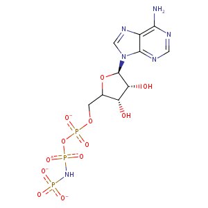
| HET Code: | ANP |
|---|---|
| Formula: | C10H13N6O12P3 |
| Molecular weight: | 502.164 g/mol |
| DrugBank ID: | - |
| Buried Surface Area: | 79.7 % |
| Polar Surface area: | 322.68 Å2 |
| Number of | |
|---|---|
| H-Bond Acceptors: | 16 |
| H-Bond Donors: | 4 |
| Rings: | 3 |
| Aromatic rings: | 2 |
| Anionic atoms: | 4 |
| Cationic atoms: | 0 |
| Rule of Five Violation: | 2 |
| Rotatable Bonds: | 8 |
| X | Y | Z |
|---|---|---|
| 37.8944 | 19.283 | 28.8162 |


Represent the protein/ligand binding mode, centered on the ligand
Dashed lines represents hydrogen bonds and metal interactions
Green residue labels for amino acids with hydrophobic contacts (green lines) to the ligand
| Ligand | Protein | Interaction | |||
|---|---|---|---|---|---|
| Atom | Atom | Residue | Distance (Å) | Angle (°) | Type |
| O2A | ND2 | ASN- 46 | 2.99 | 164.36 | H-Bond (Protein Donor) |
| N6 | OD2 | ASP- 73 | 2.59 | 145.45 | H-Bond (Ligand Donor) |
| C1' | CG1 | ILE- 78 | 4.5 | 0 | Hydrophobic |
| C5' | CB | ILE- 94 | 4 | 0 | Hydrophobic |
| C4' | CG2 | ILE- 94 | 4.32 | 0 | Hydrophobic |
| O3' | N | GLY- 102 | 2.5 | 145.81 | H-Bond (Protein Donor) |
| O1B | NZ | LYS- 103 | 2.77 | 153.47 | H-Bond (Protein Donor) |
| O1B | NZ | LYS- 103 | 2.77 | 0 | Ionic (Protein Cationic) |
| C3' | CD | LYS- 103 | 3.51 | 0 | Hydrophobic |
| C2' | CE1 | TYR- 109 | 3.72 | 0 | Hydrophobic |
| O2G | N | LEU- 115 | 2.74 | 175.56 | H-Bond (Protein Donor) |
| O2G | N | HIS- 116 | 2.69 | 166.16 | H-Bond (Protein Donor) |
| O2B | N | GLY- 117 | 2.59 | 131.02 | H-Bond (Protein Donor) |
| O3G | N | VAL- 118 | 2.63 | 146.02 | H-Bond (Protein Donor) |
| O3A | N | VAL- 118 | 3.22 | 121.28 | H-Bond (Protein Donor) |
| O3G | N | GLY- 119 | 2.73 | 164.22 | H-Bond (Protein Donor) |
| O1A | N | VAL- 120 | 3.21 | 122.3 | H-Bond (Protein Donor) |
| O1G | NZ | LYS- 337 | 3.8 | 0 | Ionic (Protein Cationic) |
| O2G | NZ | LYS- 337 | 2.65 | 0 | Ionic (Protein Cationic) |
| O2G | NZ | LYS- 337 | 2.65 | 178.18 | H-Bond (Protein Donor) |
| C1' | CD1 | ILE- 410 | 4.32 | 0 | Hydrophobic |
| O1G | O | HOH- 1560 | 2.56 | 143.05 | H-Bond (Protein Donor) |
| N1 | O | HOH- 1601 | 2.51 | 171.45 | H-Bond (Protein Donor) |
