sc-PDB
An Annotated Database of Druggable Binding Sites from the Protein DataBank
An Annotated Database of Druggable Binding Sites from the Protein DataBank
1.800 Å
X-ray
1999-12-09
| Name: | UDP-glucose 6-dehydrogenase |
|---|---|
| ID: | UDG_STRPY |
| AC: | P0C0F4 |
| Organism: | Streptococcus pyogenes |
| Reign: | Bacteria |
| TaxID: | 1314 |
| EC Number: | / |
| Chain Name: | Percentage of Residues within binding site |
|---|---|
| A | 100 % |
| B-Factor: | 24.566 |
|---|---|
| Number of residues: | 50 |
| Including | |
| Standard Amino Acids: | 43 |
| Non Standard Amino Acids: | 1 |
| Water Molecules: | 6 |
| Cofactors: | NAI |
| Metals: | |
| Ligandability | Volume (Å3) |
|---|---|
| 0.161 | 671.625 |
| % Hydrophobic | % Polar |
|---|---|
| 37.19 | 62.81 |
| According to VolSite | |
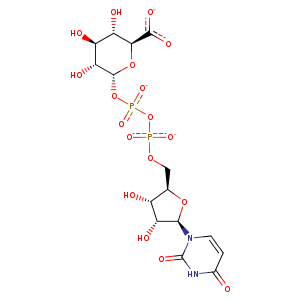
| HET Code: | UGA |
|---|---|
| Formula: | C15H19N2O18P2 |
| Molecular weight: | 577.261 g/mol |
| DrugBank ID: | DB03041 |
| Buried Surface Area: | 77.65 % |
| Polar Surface area: | 336.72 Å2 |
| Number of | |
|---|---|
| H-Bond Acceptors: | 18 |
| H-Bond Donors: | 6 |
| Rings: | 3 |
| Aromatic rings: | 0 |
| Anionic atoms: | 3 |
| Cationic atoms: | 0 |
| Rule of Five Violation: | 3 |
| Rotatable Bonds: | 9 |
| X | Y | Z |
|---|---|---|
| -14.1875 | 16.5966 | 54.9201 |
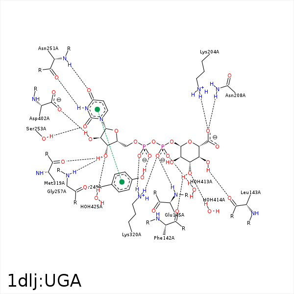


Represent the protein/ligand binding mode, centered on the ligand
Dashed lines represents hydrogen bonds and metal interactions
Green residue labels for amino acids with hydrophobic contacts (green lines) to the ligand
| Ligand | Protein | Interaction | |||
|---|---|---|---|---|---|
| Atom | Atom | Residue | Distance (Å) | Angle (°) | Type |
| O3' | O | PHE- 142 | 2.7 | 162.07 | H-Bond (Ligand Donor) |
| O4' | O | LEU- 143 | 2.65 | 168.94 | H-Bond (Ligand Donor) |
| C3' | CG | ARG- 144 | 3.92 | 0 | Hydrophobic |
| O2B | N | GLU- 145 | 2.88 | 154.41 | H-Bond (Protein Donor) |
| O'P | NZ | LYS- 204 | 2.82 | 156.44 | H-Bond (Protein Donor) |
| O'P | NZ | LYS- 204 | 2.82 | 0 | Ionic (Protein Cationic) |
| O'P | ND2 | ASN- 208 | 2.89 | 164 | H-Bond (Protein Donor) |
| C2' | CD1 | LEU- 211 | 3.79 | 0 | Hydrophobic |
| C1D | CG2 | VAL- 215 | 4.25 | 0 | Hydrophobic |
| O2A | OH | TYR- 249 | 2.69 | 177.08 | H-Bond (Protein Donor) |
| N3 | O | ASN- 251 | 2.93 | 168.71 | H-Bond (Ligand Donor) |
| O4 | N | ASN- 251 | 2.93 | 166.64 | H-Bond (Protein Donor) |
| O2 | OG | SER- 253 | 2.77 | 151.91 | H-Bond (Protein Donor) |
| C4D | CB | TYR- 256 | 4.37 | 0 | Hydrophobic |
| O3D | N | GLY- 257 | 2.88 | 139.82 | H-Bond (Protein Donor) |
| C5' | CB | SER- 260 | 4.13 | 0 | Hydrophobic |
| C1' | CD1 | LEU- 261 | 3.98 | 0 | Hydrophobic |
| C5D | CD2 | LEU- 261 | 3.99 | 0 | Hydrophobic |
| C3D | SD | MET- 319 | 3.91 | 0 | Hydrophobic |
| O3D | O | MET- 319 | 2.85 | 155.5 | H-Bond (Ligand Donor) |
| O2B | NZ | LYS- 320 | 2.83 | 163.64 | H-Bond (Protein Donor) |
| O1A | NZ | LYS- 320 | 3.23 | 156.31 | H-Bond (Protein Donor) |
| O2B | NZ | LYS- 320 | 2.83 | 0 | Ionic (Protein Cationic) |
| O1A | NZ | LYS- 320 | 3.23 | 0 | Ionic (Protein Cationic) |
| C2D | CB | LYS- 320 | 3.76 | 0 | Hydrophobic |
| O2D | OXT | ASP- 402 | 2.73 | 155.32 | H-Bond (Ligand Donor) |
| C5' | C4N | NAI- 403 | 3.74 | 0 | Hydrophobic |
| O1B | O | HOH- 413 | 2.73 | 157.98 | H-Bond (Protein Donor) |
| O3' | O | HOH- 414 | 2.85 | 179.99 | H-Bond (Protein Donor) |
| O3D | O | HOH- 425 | 3.29 | 179.96 | H-Bond (Protein Donor) |
| O4 | O | HOH- 465 | 3.06 | 120.46 | H-Bond (Protein Donor) |
