sc-PDB
An Annotated Database of Druggable Binding Sites from the Protein DataBank
An Annotated Database of Druggable Binding Sites from the Protein DataBank
1.800 Å
X-ray
1998-04-14
| Name: | UDP-glucose 4-epimerase |
|---|---|
| ID: | GALE_ECOLI |
| AC: | P09147 |
| Organism: | Escherichia coli |
| Reign: | Bacteria |
| TaxID: | 83333 |
| EC Number: | 5.1.3.2 |
| Chain Name: | Percentage of Residues within binding site |
|---|---|
| A | 100 % |
| B-Factor: | 17.945 |
|---|---|
| Number of residues: | 42 |
| Including | |
| Standard Amino Acids: | 38 |
| Non Standard Amino Acids: | 1 |
| Water Molecules: | 3 |
| Cofactors: | NAD |
| Metals: | |
| Ligandability | Volume (Å3) |
|---|---|
| 0.963 | 722.250 |
| % Hydrophobic | % Polar |
|---|---|
| 44.86 | 55.14 |
| According to VolSite | |
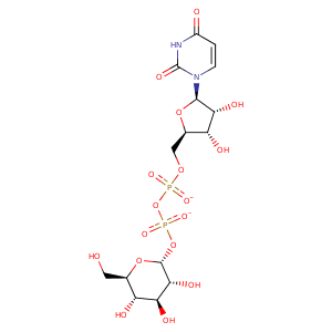
| HET Code: | UPG |
|---|---|
| Formula: | C15H22N2O17P2 |
| Molecular weight: | 564.286 g/mol |
| DrugBank ID: | DB01861 |
| Buried Surface Area: | 71.83 % |
| Polar Surface area: | 316.82 Å2 |
| Number of | |
|---|---|
| H-Bond Acceptors: | 17 |
| H-Bond Donors: | 7 |
| Rings: | 3 |
| Aromatic rings: | 0 |
| Anionic atoms: | 2 |
| Cationic atoms: | 0 |
| Rule of Five Violation: | 3 |
| Rotatable Bonds: | 9 |
| X | Y | Z |
|---|---|---|
| -23.8307 | 63.9462 | 1.37519 |
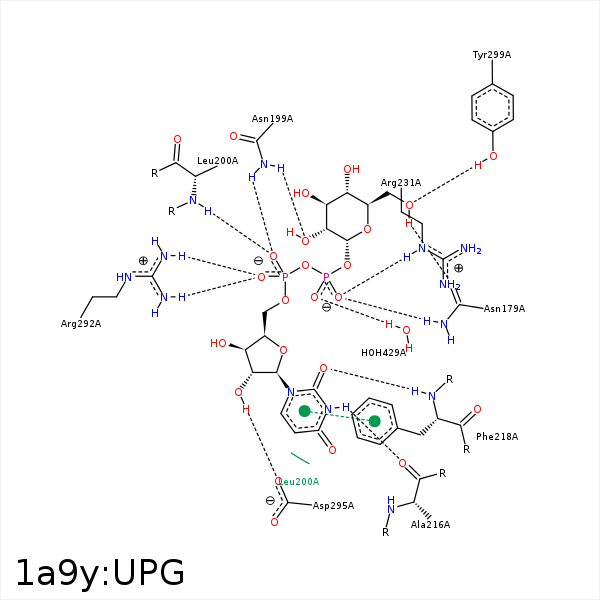


Represent the protein/ligand binding mode, centered on the ligand
Dashed lines represents hydrogen bonds and metal interactions
Green residue labels for amino acids with hydrophobic contacts (green lines) to the ligand
| Ligand | Protein | Interaction | |||
|---|---|---|---|---|---|
| Atom | Atom | Residue | Distance (Å) | Angle (°) | Type |
| C3' | CG2 | VAL- 86 | 4.46 | 0 | Hydrophobic |
| C4' | CB | ALA- 124 | 4.17 | 0 | Hydrophobic |
| C6' | CB | ALA- 125 | 3.66 | 0 | Hydrophobic |
| C3' | CE2 | PHE- 149 | 4.42 | 0 | Hydrophobic |
| O1B | ND2 | ASN- 179 | 2.83 | 163.75 | H-Bond (Protein Donor) |
| O6' | OD1 | ASN- 179 | 2.94 | 149.64 | H-Bond (Ligand Donor) |
| O2A | ND2 | ASN- 199 | 3.12 | 158.05 | H-Bond (Protein Donor) |
| O2' | ND2 | ASN- 199 | 2.87 | 154.28 | H-Bond (Protein Donor) |
| C4C | CD2 | LEU- 200 | 4.26 | 0 | Hydrophobic |
| C5C | CB | LEU- 200 | 4.17 | 0 | Hydrophobic |
| O2A | N | LEU- 200 | 2.92 | 164.65 | H-Bond (Protein Donor) |
| N3 | O | ALA- 216 | 2.8 | 170.69 | H-Bond (Ligand Donor) |
| O2 | N | PHE- 218 | 2.81 | 166.04 | H-Bond (Protein Donor) |
| C2C | CD2 | PHE- 218 | 4.45 | 0 | Hydrophobic |
| O1B | NE | ARG- 231 | 2.97 | 146.61 | H-Bond (Protein Donor) |
| C5C | CG | ARG- 231 | 3.83 | 0 | Hydrophobic |
| C5C | CZ | TYR- 233 | 4.33 | 0 | Hydrophobic |
| C1C | CG2 | VAL- 269 | 3.67 | 0 | Hydrophobic |
| C4C | CG2 | VAL- 269 | 4.22 | 0 | Hydrophobic |
| O5C | NH2 | ARG- 292 | 3.33 | 127.05 | H-Bond (Protein Donor) |
| O1A | NH2 | ARG- 292 | 2.8 | 157.98 | H-Bond (Protein Donor) |
| O1A | NH1 | ARG- 292 | 3.08 | 139.94 | H-Bond (Protein Donor) |
| O1A | CZ | ARG- 292 | 3.4 | 0 | Ionic (Protein Cationic) |
| O2C | OD2 | ASP- 295 | 2.72 | 166.63 | H-Bond (Ligand Donor) |
| C6' | CE1 | TYR- 299 | 4.49 | 0 | Hydrophobic |
| O6' | OH | TYR- 299 | 2.56 | 166.32 | H-Bond (Protein Donor) |
| C4' | C4N | NAD- 340 | 3.91 | 0 | Hydrophobic |
| O2B | O | HOH- 429 | 2.51 | 163.57 | H-Bond (Protein Donor) |
