sc-PDB
An Annotated Database of Druggable Binding Sites from the Protein DataBank
An Annotated Database of Druggable Binding Sites from the Protein DataBank
2.000 Å
X-ray
2016-03-17
| Name: | Adenosine receptor A2a |
|---|---|
| ID: | AA2AR_HUMAN |
| AC: | P29274 |
| Organism: | Homo sapiens |
| Reign: | Eukaryota |
| TaxID: | 9606 |
| EC Number: | / |
| Chain Name: | Percentage of Residues within binding site |
|---|---|
| A | 100 % |
| B-Factor: | 0.000 |
|---|---|
| Number of residues: | 26 |
| Including | |
| Standard Amino Acids: | 24 |
| Non Standard Amino Acids: | 0 |
| Water Molecules: | 2 |
| Cofactors: | |
| Metals: | |
| Ligandability | Volume (Å3) |
|---|---|
| 1.598 | 526.500 |
| % Hydrophobic | % Polar |
|---|---|
| 67.31 | 32.69 |
| According to VolSite | |
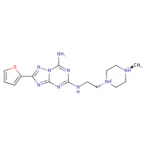
| HET Code: | 6DZ |
|---|---|
| Formula: | C15H22N9O |
| Molecular weight: | 344.395 g/mol |
| DrugBank ID: | - |
| Buried Surface Area: | 51.94 % |
| Polar Surface area: | 114.84 Å2 |
| Number of | |
|---|---|
| H-Bond Acceptors: | 7 |
| H-Bond Donors: | 3 |
| Rings: | 4 |
| Aromatic rings: | 3 |
| Anionic atoms: | 0 |
| Cationic atoms: | 1 |
| Rule of Five Violation: | 0 |
| Rotatable Bonds: | 5 |
| X | Y | Z |
|---|---|---|
| -19.9751 | 8.77388 | 16.9322 |
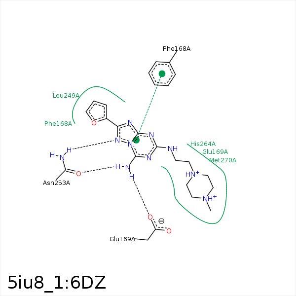


Represent the protein/ligand binding mode, centered on the ligand
Dashed lines represents hydrogen bonds and metal interactions
Green residue labels for amino acids with hydrophobic contacts (green lines) to the ligand
| Ligand | Protein | Interaction | |||
|---|---|---|---|---|---|
| Atom | Atom | Residue | Distance (Å) | Angle (°) | Type |
| C07 | CB | PHE- 168 | 3.84 | 0 | Hydrophobic |
| DuAr | DuAr | PHE- 168 | 3.69 | 0 | Aromatic Face/Face |
| N17 | OE2 | GLU- 169 | 2.67 | 176.01 | H-Bond (Ligand Donor) |
| N14 | ND2 | ASN- 253 | 3.24 | 164.35 | H-Bond (Protein Donor) |
| N17 | OD1 | ASN- 253 | 2.74 | 147.3 | H-Bond (Ligand Donor) |
