sc-PDB
An Annotated Database of Druggable Binding Sites from the Protein DataBank
An Annotated Database of Druggable Binding Sites from the Protein DataBank
2.300 Å
X-ray
2016-01-27
| Name: | Probable sugar kinase |
|---|---|
| ID: | Q31KC7_SYNE7 |
| AC: | Q31KC7 |
| Organism: | Synechococcus elongatus |
| Reign: | Bacteria |
| TaxID: | 1140 |
| EC Number: | / |
| Chain Name: | Percentage of Residues within binding site |
|---|---|
| A | 100 % |
| B-Factor: | 31.286 |
|---|---|
| Number of residues: | 29 |
| Including | |
| Standard Amino Acids: | 28 |
| Non Standard Amino Acids: | 0 |
| Water Molecules: | 1 |
| Cofactors: | |
| Metals: | |
| Ligandability | Volume (Å3) |
|---|---|
| 0.596 | 394.875 |
| % Hydrophobic | % Polar |
|---|---|
| 49.57 | 50.43 |
| According to VolSite | |
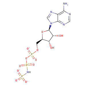
| HET Code: | ANP |
|---|---|
| Formula: | C10H13N6O12P3 |
| Molecular weight: | 502.164 g/mol |
| DrugBank ID: | - |
| Buried Surface Area: | 44.96 % |
| Polar Surface area: | 322.68 Å2 |
| Number of | |
|---|---|
| H-Bond Acceptors: | 16 |
| H-Bond Donors: | 4 |
| Rings: | 3 |
| Aromatic rings: | 2 |
| Anionic atoms: | 4 |
| Cationic atoms: | 0 |
| Rule of Five Violation: | 2 |
| Rotatable Bonds: | 8 |
| X | Y | Z |
|---|---|---|
| -13.1776 | -3.84426 | 7.28758 |
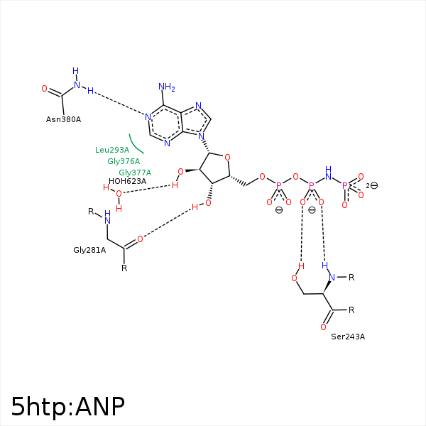


Represent the protein/ligand binding mode, centered on the ligand
Dashed lines represents hydrogen bonds and metal interactions
Green residue labels for amino acids with hydrophobic contacts (green lines) to the ligand
| Ligand | Protein | Interaction | |||
|---|---|---|---|---|---|
| Atom | Atom | Residue | Distance (Å) | Angle (°) | Type |
| O1B | OG | SER- 243 | 2.7 | 137.83 | H-Bond (Protein Donor) |
| O2B | N | SER- 243 | 2.79 | 165.23 | H-Bond (Protein Donor) |
| O3' | O | GLY- 281 | 2.95 | 139.21 | H-Bond (Ligand Donor) |
| C2' | CD1 | LEU- 293 | 4.36 | 0 | Hydrophobic |
| O2A | N | GLY- 376 | 3.23 | 138.47 | H-Bond (Protein Donor) |
| N1 | ND2 | ASN- 380 | 3.09 | 159.65 | H-Bond (Protein Donor) |
| O2' | O | HOH- 623 | 2.84 | 168.38 | H-Bond (Ligand Donor) |
