sc-PDB
An Annotated Database of Druggable Binding Sites from the Protein DataBank
An Annotated Database of Druggable Binding Sites from the Protein DataBank
1.800 Å
X-ray
2016-05-01
| Name: | Flavin-dependent L-tryptophan oxidase VioA |
|---|---|
| ID: | VIOA_CHRVO |
| AC: | Q9S3V1 |
| Organism: | Chromobacterium violaceum |
| Reign: | Bacteria |
| TaxID: | 243365 |
| EC Number: | / |
| Chain Name: | Percentage of Residues within binding site |
|---|---|
| C | 100 % |
| B-Factor: | 16.567 |
|---|---|
| Number of residues: | 66 |
| Including | |
| Standard Amino Acids: | 62 |
| Non Standard Amino Acids: | 1 |
| Water Molecules: | 3 |
| Cofactors: | |
| Metals: | CL |
| Ligandability | Volume (Å3) |
|---|---|
| 1.084 | 1599.750 |
| % Hydrophobic | % Polar |
|---|---|
| 50.84 | 49.16 |
| According to VolSite | |
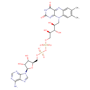
| HET Code: | FDA |
|---|---|
| Formula: | C27H33N9O15P2 |
| Molecular weight: | 785.550 g/mol |
| DrugBank ID: | - |
| Buried Surface Area: | 75.98 % |
| Polar Surface area: | 381.04 Å2 |
| Number of | |
|---|---|
| H-Bond Acceptors: | 21 |
| H-Bond Donors: | 9 |
| Rings: | 6 |
| Aromatic rings: | 3 |
| Anionic atoms: | 2 |
| Cationic atoms: | 0 |
| Rule of Five Violation: | 3 |
| Rotatable Bonds: | 13 |
| X | Y | Z |
|---|---|---|
| 64.3346 | -48.8167 | 108.854 |
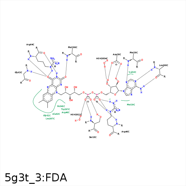


Represent the protein/ligand binding mode, centered on the ligand
Dashed lines represents hydrogen bonds and metal interactions
Green residue labels for amino acids with hydrophobic contacts (green lines) to the ligand
| Ligand | Protein | Interaction | |||
|---|---|---|---|---|---|
| Atom | Atom | Residue | Distance (Å) | Angle (°) | Type |
| C4' | CG2 | ILE- 14 | 4.31 | 0 | Hydrophobic |
| O1P | N | SER- 15 | 2.92 | 161.54 | H-Bond (Protein Donor) |
| O2P | OG | SER- 15 | 2.75 | 169.34 | H-Bond (Protein Donor) |
| O3B | OD2 | ASP- 38 | 2.6 | 166.84 | H-Bond (Ligand Donor) |
| O3B | OD1 | ASP- 38 | 3.21 | 129.15 | H-Bond (Ligand Donor) |
| O2B | OD1 | ASP- 38 | 2.64 | 157.18 | H-Bond (Ligand Donor) |
| C2B | CE | MET- 39 | 3.96 | 0 | Hydrophobic |
| C1B | CG | MET- 39 | 4.35 | 0 | Hydrophobic |
| N3A | N | MET- 39 | 3.11 | 142.95 | H-Bond (Protein Donor) |
| O1A | N | ARG- 46 | 2.9 | 169.62 | H-Bond (Protein Donor) |
| O2A | NH2 | ARG- 46 | 3.15 | 137.69 | H-Bond (Protein Donor) |
| O2A | NE | ARG- 46 | 2.81 | 160.69 | H-Bond (Protein Donor) |
| O3P | NH2 | ARG- 46 | 3.39 | 145.13 | H-Bond (Protein Donor) |
| O2A | CZ | ARG- 46 | 3.42 | 0 | Ionic (Protein Cationic) |
| C8M | CD | ARG- 46 | 3.95 | 0 | Hydrophobic |
| C9 | CB | ARG- 46 | 4.34 | 0 | Hydrophobic |
| C9A | CB | ALA- 62 | 4.07 | 0 | Hydrophobic |
| C2' | CB | ALA- 62 | 4.03 | 0 | Hydrophobic |
| O4 | N | GLY- 63 | 3.09 | 160.31 | H-Bond (Protein Donor) |
| N3 | O | ARG- 64 | 2.84 | 164.94 | H-Bond (Ligand Donor) |
| O4 | N | ARG- 64 | 3.01 | 143.81 | H-Bond (Protein Donor) |
| O4 | NE | ARG- 64 | 2.97 | 127.57 | H-Bond (Protein Donor) |
| N6A | O | LEU- 208 | 3.04 | 167.29 | H-Bond (Ligand Donor) |
| N1A | N | LEU- 208 | 2.86 | 153.2 | H-Bond (Protein Donor) |
| C1B | CG2 | ILE- 241 | 4.17 | 0 | Hydrophobic |
| C7 | CD1 | LEU- 267 | 3.67 | 0 | Hydrophobic |
| C8 | CD1 | LEU- 267 | 3.7 | 0 | Hydrophobic |
| C7M | CD | LYS- 269 | 4.21 | 0 | Hydrophobic |
| C7M | CE1 | TYR- 309 | 4.07 | 0 | Hydrophobic |
| C8M | CD2 | TRP- 359 | 3.7 | 0 | Hydrophobic |
| N1 | N | MET- 398 | 3.37 | 133.84 | H-Bond (Protein Donor) |
| O2 | N | MET- 398 | 2.81 | 168.59 | H-Bond (Protein Donor) |
| C2' | CG | MET- 398 | 3.97 | 0 | Hydrophobic |
| C4' | CG | MET- 398 | 4.33 | 0 | Hydrophobic |
| O1P | O | HOH- 2011 | 2.78 | 162.5 | H-Bond (Protein Donor) |
| O3B | O | HOH- 2054 | 2.82 | 135.01 | H-Bond (Protein Donor) |
