sc-PDB
An Annotated Database of Druggable Binding Sites from the Protein DataBank
An Annotated Database of Druggable Binding Sites from the Protein DataBank
2.660 Å
X-ray
2015-09-17
| Name: | Erythronate-4-phosphate dehydrogenase |
|---|---|
| ID: | PDXB_VIBCH |
| AC: | Q9KQ92 |
| Organism: | Vibrio cholerae serotype O1 |
| Reign: | Bacteria |
| TaxID: | 243277 |
| EC Number: | / |
| Chain Name: | Percentage of Residues within binding site |
|---|---|
| A | 100 % |
| B-Factor: | 55.685 |
|---|---|
| Number of residues: | 45 |
| Including | |
| Standard Amino Acids: | 44 |
| Non Standard Amino Acids: | 1 |
| Water Molecules: | 0 |
| Cofactors: | |
| Metals: | CL |
| Ligandability | Volume (Å3) |
|---|---|
| 0.241 | 499.500 |
| % Hydrophobic | % Polar |
|---|---|
| 37.84 | 62.16 |
| According to VolSite | |
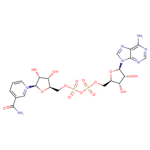
| HET Code: | NAD |
|---|---|
| Formula: | C21H26N7O14P2 |
| Molecular weight: | 662.417 g/mol |
| DrugBank ID: | - |
| Buried Surface Area: | 62.66 % |
| Polar Surface area: | 343.54 Å2 |
| Number of | |
|---|---|
| H-Bond Acceptors: | 18 |
| H-Bond Donors: | 6 |
| Rings: | 5 |
| Aromatic rings: | 3 |
| Anionic atoms: | 2 |
| Cationic atoms: | 1 |
| Rule of Five Violation: | 3 |
| Rotatable Bonds: | 11 |
| X | Y | Z |
|---|---|---|
| 20.2742 | 51.0925 | 18.2601 |
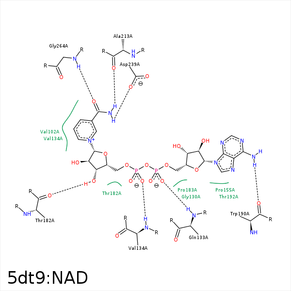


Represent the protein/ligand binding mode, centered on the ligand
Dashed lines represents hydrogen bonds and metal interactions
Green residue labels for amino acids with hydrophobic contacts (green lines) to the ligand
| Ligand | Protein | Interaction | |||
|---|---|---|---|---|---|
| Atom | Atom | Residue | Distance (Å) | Angle (°) | Type |
| C4N | CG2 | VAL- 102 | 3.87 | 0 | Hydrophobic |
| O2A | N | GLN- 133 | 3.09 | 161.85 | H-Bond (Protein Donor) |
| O2N | N | VAL- 134 | 2.93 | 171.98 | H-Bond (Protein Donor) |
| C5D | CG2 | VAL- 134 | 3.76 | 0 | Hydrophobic |
| C3B | CG | LYS- 156 | 4.46 | 0 | Hydrophobic |
| C5D | CB | HIS- 181 | 4.39 | 0 | Hydrophobic |
| O3D | O | THR- 182 | 2.78 | 159.51 | H-Bond (Ligand Donor) |
| N6A | O | TRP- 190 | 3 | 139.99 | H-Bond (Ligand Donor) |
| N7N | O | ALA- 213 | 2.62 | 123.58 | H-Bond (Ligand Donor) |
| C3D | CD | ARG- 215 | 4.4 | 0 | Hydrophobic |
| N7N | OD2 | ASP- 239 | 3.02 | 168.13 | H-Bond (Ligand Donor) |
| O7N | N | GLY- 264 | 2.77 | 149.72 | H-Bond (Protein Donor) |
