sc-PDB
An Annotated Database of Druggable Binding Sites from the Protein DataBank
An Annotated Database of Druggable Binding Sites from the Protein DataBank
2.590 Å
X-ray
2015-06-26
| Name: | Tubulin tyrosine ligase |
|---|---|
| ID: | E1BQ43_CHICK |
| AC: | E1BQ43 |
| Organism: | Gallus gallus |
| Reign: | Eukaryota |
| TaxID: | 9031 |
| EC Number: | / |
| Chain Name: | Percentage of Residues within binding site |
|---|---|
| F | 100 % |
| B-Factor: | 87.760 |
|---|---|
| Number of residues: | 26 |
| Including | |
| Standard Amino Acids: | 26 |
| Non Standard Amino Acids: | 0 |
| Water Molecules: | 0 |
| Cofactors: | |
| Metals: | |
| Ligandability | Volume (Å3) |
|---|---|
| 0.882 | 651.375 |
| % Hydrophobic | % Polar |
|---|---|
| 50.26 | 49.74 |
| According to VolSite | |
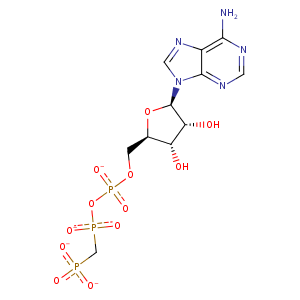
| HET Code: | ACP |
|---|---|
| Formula: | C11H14N5O12P3 |
| Molecular weight: | 501.176 g/mol |
| DrugBank ID: | DB03909 |
| Buried Surface Area: | 59.99 % |
| Polar Surface area: | 310.64 Å2 |
| Number of | |
|---|---|
| H-Bond Acceptors: | 16 |
| H-Bond Donors: | 3 |
| Rings: | 3 |
| Aromatic rings: | 2 |
| Anionic atoms: | 4 |
| Cationic atoms: | 0 |
| Rule of Five Violation: | 2 |
| Rotatable Bonds: | 8 |
| X | Y | Z |
|---|---|---|
| -8.65865 | -50.7017 | 100.077 |
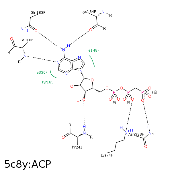


Represent the protein/ligand binding mode, centered on the ligand
Dashed lines represents hydrogen bonds and metal interactions
Green residue labels for amino acids with hydrophobic contacts (green lines) to the ligand
| Ligand | Protein | Interaction | |||
|---|---|---|---|---|---|
| Atom | Atom | Residue | Distance (Å) | Angle (°) | Type |
| O1B | NZ | LYS- 74 | 3.9 | 0 | Ionic (Protein Cationic) |
| O2B | NZ | LYS- 74 | 3.09 | 0 | Ionic (Protein Cationic) |
| O2A | NZ | LYS- 74 | 3.5 | 0 | Ionic (Protein Cationic) |
| O2B | NZ | LYS- 74 | 3.09 | 163.63 | H-Bond (Protein Donor) |
| N6 | OE1 | GLN- 183 | 2.9 | 169.49 | H-Bond (Ligand Donor) |
| N6 | O | LYS- 184 | 3.15 | 147.52 | H-Bond (Ligand Donor) |
| N1 | N | LEU- 186 | 3.34 | 159.88 | H-Bond (Protein Donor) |
| C4' | CD2 | LEU- 240 | 4.33 | 0 | Hydrophobic |
| C1' | CD2 | LEU- 240 | 4.39 | 0 | Hydrophobic |
| O3' | N | THR- 241 | 3.14 | 152.42 | H-Bond (Protein Donor) |
| O1G | ND2 | ASN- 333 | 2.59 | 155.62 | H-Bond (Protein Donor) |
