sc-PDB
An Annotated Database of Druggable Binding Sites from the Protein DataBank
An Annotated Database of Druggable Binding Sites from the Protein DataBank
2.500 Å
X-ray
2015-06-24
| Name: | Protein arginine N-methyltransferase SFM1 |
|---|---|
| ID: | SFM1_YEAST |
| AC: | Q12314 |
| Organism: | Saccharomyces cerevisiae |
| Reign: | Eukaryota |
| TaxID: | 559292 |
| EC Number: | / |
| Chain Name: | Percentage of Residues within binding site |
|---|---|
| A | 100 % |
| B-Factor: | 44.006 |
|---|---|
| Number of residues: | 30 |
| Including | |
| Standard Amino Acids: | 29 |
| Non Standard Amino Acids: | 0 |
| Water Molecules: | 1 |
| Cofactors: | |
| Metals: | |
| Ligandability | Volume (Å3) |
|---|---|
| 0.752 | 654.750 |
| % Hydrophobic | % Polar |
|---|---|
| 40.72 | 59.28 |
| According to VolSite | |
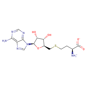
| HET Code: | SAH |
|---|---|
| Formula: | C14H20N6O5S |
| Molecular weight: | 384.411 g/mol |
| DrugBank ID: | DB01752 |
| Buried Surface Area: | 64.16 % |
| Polar Surface area: | 212.38 Å2 |
| Number of | |
|---|---|
| H-Bond Acceptors: | 10 |
| H-Bond Donors: | 4 |
| Rings: | 3 |
| Aromatic rings: | 2 |
| Anionic atoms: | 1 |
| Cationic atoms: | 1 |
| Rule of Five Violation: | 1 |
| Rotatable Bonds: | 7 |
| X | Y | Z |
|---|---|---|
| -16.9283 | -17.2117 | 86.1467 |
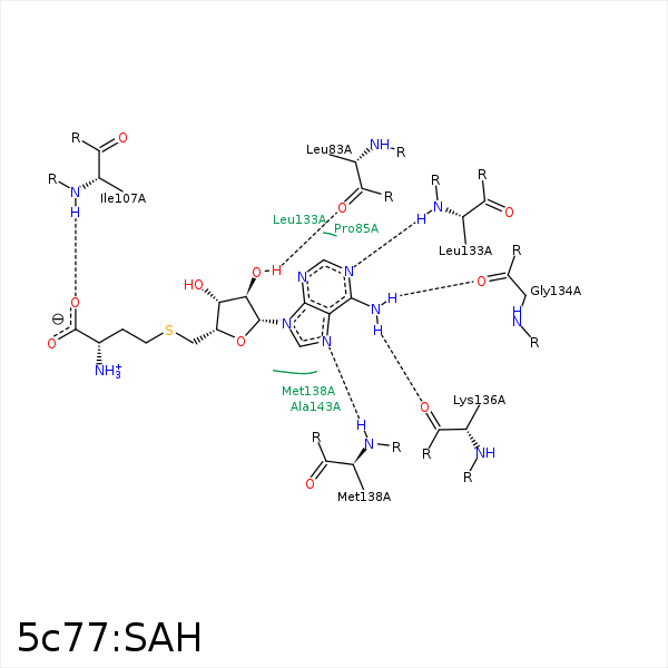


Represent the protein/ligand binding mode, centered on the ligand
Dashed lines represents hydrogen bonds and metal interactions
Green residue labels for amino acids with hydrophobic contacts (green lines) to the ligand
| Ligand | Protein | Interaction | |||
|---|---|---|---|---|---|
| Atom | Atom | Residue | Distance (Å) | Angle (°) | Type |
| O2' | O | LEU- 83 | 2.78 | 169.1 | H-Bond (Ligand Donor) |
| C1' | CD2 | PHE- 104 | 4.1 | 0 | Hydrophobic |
| O | N | ILE- 107 | 2.89 | 164.39 | H-Bond (Protein Donor) |
| N1 | N | LEU- 133 | 2.92 | 148.99 | H-Bond (Protein Donor) |
| N6 | O | GLY- 134 | 3.27 | 167.13 | H-Bond (Ligand Donor) |
| N6 | O | LYS- 136 | 2.72 | 133.07 | H-Bond (Ligand Donor) |
| N7 | N | MET- 138 | 2.99 | 147.16 | H-Bond (Protein Donor) |
| SD | CB | THR- 140 | 4.44 | 0 | Hydrophobic |
| C1' | CB | ALA- 143 | 4.01 | 0 | Hydrophobic |
