sc-PDB
An Annotated Database of Druggable Binding Sites from the Protein DataBank
An Annotated Database of Druggable Binding Sites from the Protein DataBank
2.500 Å
X-ray
2015-06-08
| Name: | ADP-ribosyltransferase |
|---|---|
| ID: | Q8KNY0_BACCE |
| AC: | Q8KNY0 |
| Organism: | Bacillus cereus |
| Reign: | Bacteria |
| TaxID: | 1396 |
| EC Number: | / |
| Chain Name: | Percentage of Residues within binding site |
|---|---|
| A | 9 % |
| B | 91 % |
| B-Factor: | 71.665 |
|---|---|
| Number of residues: | 45 |
| Including | |
| Standard Amino Acids: | 44 |
| Non Standard Amino Acids: | 0 |
| Water Molecules: | 1 |
| Cofactors: | |
| Metals: | |
| Ligandability | Volume (Å3) |
|---|---|
| 0.274 | 958.500 |
| % Hydrophobic | % Polar |
|---|---|
| 37.32 | 62.68 |
| According to VolSite | |
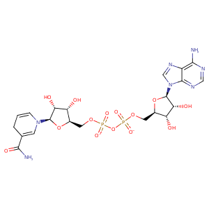
| HET Code: | NAI |
|---|---|
| Formula: | C21H27N7O14P2 |
| Molecular weight: | 663.425 g/mol |
| DrugBank ID: | DB00157 |
| Buried Surface Area: | 66.09 % |
| Polar Surface area: | 342.9 Å2 |
| Number of | |
|---|---|
| H-Bond Acceptors: | 19 |
| H-Bond Donors: | 6 |
| Rings: | 5 |
| Aromatic rings: | 2 |
| Anionic atoms: | 2 |
| Cationic atoms: | 0 |
| Rule of Five Violation: | 3 |
| Rotatable Bonds: | 11 |
| X | Y | Z |
|---|---|---|
| -15.999 | 17.6654 | 13.5735 |
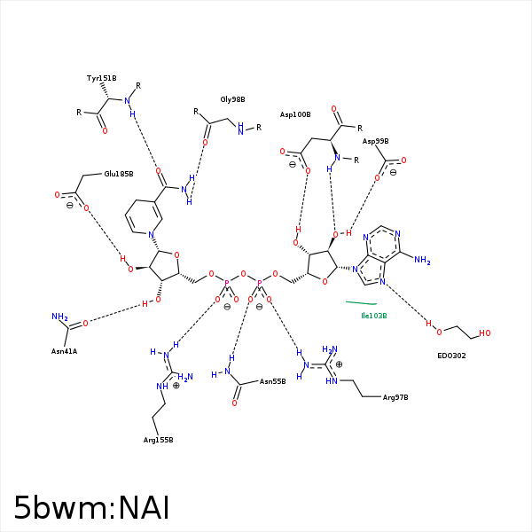


Represent the protein/ligand binding mode, centered on the ligand
Dashed lines represents hydrogen bonds and metal interactions
Green residue labels for amino acids with hydrophobic contacts (green lines) to the ligand
| Ligand | Protein | Interaction | |||
|---|---|---|---|---|---|
| Atom | Atom | Residue | Distance (Å) | Angle (°) | Type |
| O3D | OD1 | ASN- 41 | 3.2 | 165.06 | H-Bond (Ligand Donor) |
| C3D | CE2 | TYR- 47 | 4.21 | 0 | Hydrophobic |
| C3D | CB | ALA- 51 | 3.69 | 0 | Hydrophobic |
| O1A | ND2 | ASN- 55 | 2.88 | 161.63 | H-Bond (Protein Donor) |
| O2A | CZ | ARG- 97 | 3.4 | 0 | Ionic (Protein Cationic) |
| O7N | N | GLY- 98 | 3.31 | 149.65 | H-Bond (Protein Donor) |
| N7N | O | GLY- 98 | 3.29 | 160.63 | H-Bond (Ligand Donor) |
| O2B | OD1 | ASP- 99 | 3.08 | 148.65 | H-Bond (Ligand Donor) |
| O2B | N | ASP- 100 | 2.98 | 165.98 | H-Bond (Protein Donor) |
| O3B | NE1 | TRP- 102 | 3.4 | 121.3 | H-Bond (Protein Donor) |
| C4N | CB | SER- 142 | 4.36 | 0 | Hydrophobic |
| C2D | CB | SER- 142 | 3.48 | 0 | Hydrophobic |
| C4N | CB | SER- 144 | 4.32 | 0 | Hydrophobic |
| C1D | CE2 | TYR- 151 | 4.15 | 0 | Hydrophobic |
| O7N | N | TYR- 151 | 2.69 | 154.81 | H-Bond (Protein Donor) |
| O1N | NH1 | ARG- 155 | 2.63 | 171.98 | H-Bond (Protein Donor) |
| O2N | NH2 | ARG- 155 | 3.36 | 159.97 | H-Bond (Protein Donor) |
| O1N | CZ | ARG- 155 | 3.53 | 0 | Ionic (Protein Cationic) |
| O2D | OE2 | GLU- 185 | 3.37 | 128.53 | H-Bond (Ligand Donor) |
| O2D | OE1 | GLU- 185 | 2.83 | 170.24 | H-Bond (Ligand Donor) |
