sc-PDB
An Annotated Database of Druggable Binding Sites from the Protein DataBank
An Annotated Database of Druggable Binding Sites from the Protein DataBank
2.100 Å
X-ray
2015-02-05
| Name: | Leucine--tRNA ligase |
|---|---|
| ID: | B9JQP8_AGRRK |
| AC: | B9JQP8 |
| Organism: | Agrobacterium radiobacter |
| Reign: | Bacteria |
| TaxID: | 311403 |
| EC Number: | / |
| Chain Name: | Percentage of Residues within binding site |
|---|---|
| A | 100 % |
| B-Factor: | 31.828 |
|---|---|
| Number of residues: | 38 |
| Including | |
| Standard Amino Acids: | 36 |
| Non Standard Amino Acids: | 0 |
| Water Molecules: | 2 |
| Cofactors: | |
| Metals: | |
| Ligandability | Volume (Å3) |
|---|---|
| 0.764 | 1181.250 |
| % Hydrophobic | % Polar |
|---|---|
| 31.43 | 68.57 |
| According to VolSite | |
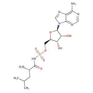
| HET Code: | LSS |
|---|---|
| Formula: | C16H25N7O7S |
| Molecular weight: | 459.477 g/mol |
| DrugBank ID: | - |
| Buried Surface Area: | 74.17 % |
| Polar Surface area: | 229.86 Å2 |
| Number of | |
|---|---|
| H-Bond Acceptors: | 11 |
| H-Bond Donors: | 4 |
| Rings: | 3 |
| Aromatic rings: | 2 |
| Anionic atoms: | 1 |
| Cationic atoms: | 1 |
| Rule of Five Violation: | 1 |
| Rotatable Bonds: | 8 |
| X | Y | Z |
|---|---|---|
| 19.4805 | -1.10768 | 2.43216 |
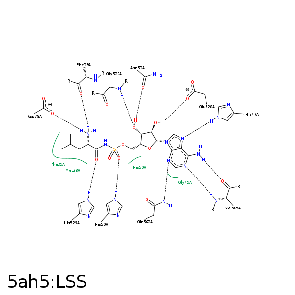


Represent the protein/ligand binding mode, centered on the ligand
Dashed lines represents hydrogen bonds and metal interactions
Green residue labels for amino acids with hydrophobic contacts (green lines) to the ligand
| Ligand | Protein | Interaction | |||
|---|---|---|---|---|---|
| Atom | Atom | Residue | Distance (Å) | Angle (°) | Type |
| C23 | CB | MET- 38 | 4.02 | 0 | Hydrophobic |
| C11 | CB | MET- 38 | 3.73 | 0 | Hydrophobic |
| N4 | O | PHE- 39 | 2.88 | 158.1 | H-Bond (Ligand Donor) |
| C7 | CE1 | PHE- 39 | 3.81 | 0 | Hydrophobic |
| C9 | CD1 | PHE- 39 | 4.49 | 0 | Hydrophobic |
| C11 | CD1 | PHE- 39 | 3.79 | 0 | Hydrophobic |
| O2A | N | TYR- 41 | 3.35 | 143.36 | H-Bond (Protein Donor) |
| N7 | NE2 | HIS- 47 | 2.84 | 168.75 | H-Bond (Protein Donor) |
| O2A | NE2 | HIS- 50 | 2.82 | 161.83 | H-Bond (Protein Donor) |
| O3 | OD1 | ASN- 53 | 2.52 | 158.93 | H-Bond (Ligand Donor) |
| C21 | CB | ASN- 53 | 4.38 | 0 | Hydrophobic |
| N4 | OD2 | ASP- 78 | 2.71 | 154.53 | H-Bond (Ligand Donor) |
| N4 | OD2 | ASP- 78 | 2.71 | 0 | Ionic (Ligand Cationic) |
| C10 | CE1 | PHE- 490 | 4.14 | 0 | Hydrophobic |
| C10 | CB | SER- 493 | 3.71 | 0 | Hydrophobic |
| C11 | CE1 | TYR- 496 | 4.11 | 0 | Hydrophobic |
| O3 | N | GLY- 526 | 3.2 | 138.7 | H-Bond (Protein Donor) |
| O1 | NE2 | HIS- 529 | 2.79 | 162.57 | H-Bond (Protein Donor) |
| C10 | CB | HIS- 533 | 3.58 | 0 | Hydrophobic |
| C11 | CB | HIS- 533 | 4.49 | 0 | Hydrophobic |
| O2 | NE2 | GLN- 562 | 3.32 | 129.85 | H-Bond (Protein Donor) |
| N3 | NE2 | GLN- 562 | 3.01 | 160.99 | H-Bond (Protein Donor) |
| N1 | N | VAL- 565 | 2.87 | 174.62 | H-Bond (Protein Donor) |
| N6 | O | VAL- 565 | 3.23 | 173.07 | H-Bond (Ligand Donor) |
