sc-PDB
An Annotated Database of Druggable Binding Sites from the Protein DataBank
An Annotated Database of Druggable Binding Sites from the Protein DataBank
1.240 Å
X-ray
2015-08-17
| Name: | 3-phosphoinositide-dependent protein kinase 1 |
|---|---|
| ID: | PDPK1_HUMAN |
| AC: | O15530 |
| Organism: | Homo sapiens |
| Reign: | Eukaryota |
| TaxID: | 9606 |
| EC Number: | 2.7.11.1 |
| Chain Name: | Percentage of Residues within binding site |
|---|---|
| A | 100 % |
| B-Factor: | 21.649 |
|---|---|
| Number of residues: | 32 |
| Including | |
| Standard Amino Acids: | 28 |
| Non Standard Amino Acids: | 3 |
| Water Molecules: | 1 |
| Cofactors: | |
| Metals: | NA NA NA |
| Ligandability | Volume (Å3) |
|---|---|
| 0.791 | 533.250 |
| % Hydrophobic | % Polar |
|---|---|
| 44.30 | 55.70 |
| According to VolSite | |
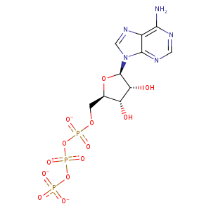
| HET Code: | ATP |
|---|---|
| Formula: | C10H12N5O13P3 |
| Molecular weight: | 503.149 g/mol |
| DrugBank ID: | DB00171 |
| Buried Surface Area: | 54.14 % |
| Polar Surface area: | 319.88 Å2 |
| Number of | |
|---|---|
| H-Bond Acceptors: | 17 |
| H-Bond Donors: | 3 |
| Rings: | 3 |
| Aromatic rings: | 2 |
| Anionic atoms: | 4 |
| Cationic atoms: | 0 |
| Rule of Five Violation: | 2 |
| Rotatable Bonds: | 8 |
| X | Y | Z |
|---|---|---|
| 51.1761 | -1.4659 | 14.3912 |
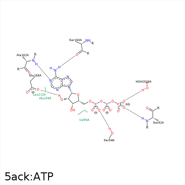


Represent the protein/ligand binding mode, centered on the ligand
Dashed lines represents hydrogen bonds and metal interactions
Green residue labels for amino acids with hydrophobic contacts (green lines) to the ligand
| Ligand | Protein | Interaction | |||
|---|---|---|---|---|---|
| Atom | Atom | Residue | Distance (Å) | Angle (°) | Type |
| O2G | N | SER- 92 | 3.04 | 174.58 | H-Bond (Protein Donor) |
| O2A | OG | SER- 94 | 2.59 | 137.6 | H-Bond (Protein Donor) |
| C5' | CG2 | VAL- 96 | 3.87 | 0 | Hydrophobic |
| O1A | NZ | LYS- 111 | 3.74 | 0 | Ionic (Protein Cationic) |
| N6 | O | SER- 160 | 2.76 | 163.45 | H-Bond (Ligand Donor) |
| N1 | N | ALA- 162 | 2.94 | 174.32 | H-Bond (Protein Donor) |
| O2' | OE2 | GLU- 166 | 2.88 | 153.13 | H-Bond (Ligand Donor) |
| O3G | O | HOH- 2020 | 2.81 | 179.96 | H-Bond (Protein Donor) |
