sc-PDB
An Annotated Database of Druggable Binding Sites from the Protein DataBank
An Annotated Database of Druggable Binding Sites from the Protein DataBank
2.470 Å
X-ray
2015-04-14
| Name: | CalS13 |
|---|---|
| ID: | Q8KND8_MICEC |
| AC: | Q8KND8 |
| Organism: | Micromonospora echinospora |
| Reign: | Bacteria |
| TaxID: | 1877 |
| EC Number: | / |
| Chain Name: | Percentage of Residues within binding site |
|---|---|
| E | 50 % |
| F | 50 % |
| B-Factor: | 67.629 |
|---|---|
| Number of residues: | 22 |
| Including | |
| Standard Amino Acids: | 21 |
| Non Standard Amino Acids: | 1 |
| Water Molecules: | 0 |
| Cofactors: | |
| Metals: | |
| Ligandability | Volume (Å3) |
|---|---|
| 0.986 | 1042.875 |
| % Hydrophobic | % Polar |
|---|---|
| 52.10 | 47.90 |
| According to VolSite | |
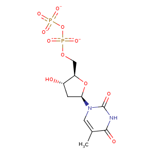
| HET Code: | TYD |
|---|---|
| Formula: | C10H13N2O11P2 |
| Molecular weight: | 399.165 g/mol |
| DrugBank ID: | DB03103 |
| Buried Surface Area: | 47.23 % |
| Polar Surface area: | 220.27 Å2 |
| Number of | |
|---|---|
| H-Bond Acceptors: | 11 |
| H-Bond Donors: | 2 |
| Rings: | 2 |
| Aromatic rings: | 0 |
| Anionic atoms: | 3 |
| Cationic atoms: | 0 |
| Rule of Five Violation: | 1 |
| Rotatable Bonds: | 6 |
| X | Y | Z |
|---|---|---|
| 14.6832 | 49.29 | 29.4871 |
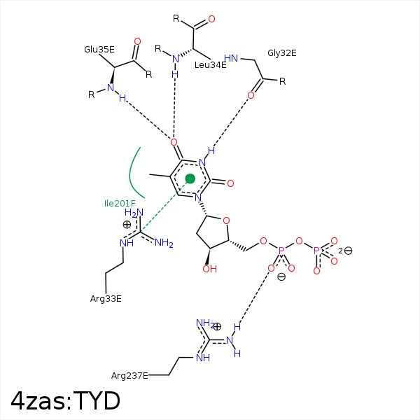


Represent the protein/ligand binding mode, centered on the ligand
Dashed lines represents hydrogen bonds and metal interactions
Green residue labels for amino acids with hydrophobic contacts (green lines) to the ligand
| Ligand | Protein | Interaction | |||
|---|---|---|---|---|---|
| Atom | Atom | Residue | Distance (Å) | Angle (°) | Type |
| C4' | CB | SER- 13 | 4.39 | 0 | Hydrophobic |
| N3 | O | GLY- 32 | 2.64 | 150.11 | H-Bond (Ligand Donor) |
| O4 | N | LEU- 34 | 2.7 | 150.75 | H-Bond (Protein Donor) |
| O4 | N | GLU- 35 | 3.19 | 170.44 | H-Bond (Protein Donor) |
| C5M | CB | GLU- 35 | 4.12 | 0 | Hydrophobic |
| C5M | CB | ALA- 200 | 4.22 | 0 | Hydrophobic |
| C1' | CG2 | ILE- 201 | 4.27 | 0 | Hydrophobic |
| C5' | CD1 | ILE- 201 | 3.9 | 0 | Hydrophobic |
| O1A | CZ | ARG- 237 | 3.92 | 0 | Ionic (Protein Cationic) |
| O1A | NH1 | ARG- 237 | 2.85 | 128.66 | H-Bond (Protein Donor) |
