sc-PDB
An Annotated Database of Druggable Binding Sites from the Protein DataBank
An Annotated Database of Druggable Binding Sites from the Protein DataBank
1.780 Å
X-ray
2015-03-11
| Name: | Glucose oxidase, putative |
|---|---|
| ID: | B8MX95_ASPFN |
| AC: | B8MX95 |
| Organism: | Aspergillus flavus |
| Reign: | Eukaryota |
| TaxID: | 332952 |
| EC Number: | / |
| Chain Name: | Percentage of Residues within binding site |
|---|---|
| A | 100 % |
| B-Factor: | 18.947 |
|---|---|
| Number of residues: | 65 |
| Including | |
| Standard Amino Acids: | 61 |
| Non Standard Amino Acids: | 0 |
| Water Molecules: | 4 |
| Cofactors: | |
| Metals: | |
| Ligandability | Volume (Å3) |
|---|---|
| 1.255 | 837.000 |
| % Hydrophobic | % Polar |
|---|---|
| 53.63 | 46.37 |
| According to VolSite | |
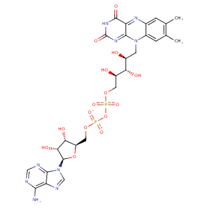
| HET Code: | FDA |
|---|---|
| Formula: | C27H33N9O15P2 |
| Molecular weight: | 785.550 g/mol |
| DrugBank ID: | - |
| Buried Surface Area: | 80.02 % |
| Polar Surface area: | 381.04 Å2 |
| Number of | |
|---|---|
| H-Bond Acceptors: | 21 |
| H-Bond Donors: | 9 |
| Rings: | 6 |
| Aromatic rings: | 3 |
| Anionic atoms: | 2 |
| Cationic atoms: | 0 |
| Rule of Five Violation: | 3 |
| Rotatable Bonds: | 13 |
| X | Y | Z |
|---|---|---|
| 27.7487 | 39.5325 | 54.1324 |


Represent the protein/ligand binding mode, centered on the ligand
Dashed lines represents hydrogen bonds and metal interactions
Green residue labels for amino acids with hydrophobic contacts (green lines) to the ligand
| Ligand | Protein | Interaction | |||
|---|---|---|---|---|---|
| Atom | Atom | Residue | Distance (Å) | Angle (°) | Type |
| O2A | OG1 | THR- 15 | 2.66 | 162.26 | H-Bond (Protein Donor) |
| O2A | N | THR- 15 | 3.17 | 168.2 | H-Bond (Protein Donor) |
| O4' | OG1 | THR- 15 | 2.77 | 165.13 | H-Bond (Ligand Donor) |
| C4' | CB | THR- 15 | 4.25 | 0 | Hydrophobic |
| O1P | N | SER- 16 | 3.03 | 147.97 | H-Bond (Protein Donor) |
| O1P | OG | SER- 16 | 2.8 | 159.83 | H-Bond (Protein Donor) |
| O3B | OE1 | GLU- 36 | 2.68 | 165.02 | H-Bond (Ligand Donor) |
| O2B | OE2 | GLU- 36 | 2.73 | 140.48 | H-Bond (Ligand Donor) |
| N3A | N | ALA- 37 | 3.25 | 138.13 | H-Bond (Protein Donor) |
| C7M | CE1 | TYR- 53 | 4.09 | 0 | Hydrophobic |
| C7M | CZ | PHE- 57 | 3.48 | 0 | Hydrophobic |
| C2B | CE2 | TRP- 63 | 4.33 | 0 | Hydrophobic |
| O2B | NE1 | TRP- 63 | 3.17 | 145.4 | H-Bond (Protein Donor) |
| C8M | CB | ARG- 81 | 4.06 | 0 | Hydrophobic |
| O1A | OG1 | THR- 89 | 2.64 | 169.07 | H-Bond (Protein Donor) |
| O2A | N | THR- 89 | 2.9 | 167.73 | H-Bond (Protein Donor) |
| C8M | CG2 | THR- 89 | 3.52 | 0 | Hydrophobic |
| C9 | CG2 | THR- 89 | 3.55 | 0 | Hydrophobic |
| C3' | CG2 | THR- 89 | 3.93 | 0 | Hydrophobic |
| C4' | CB | THR- 89 | 4.12 | 0 | Hydrophobic |
| C8M | CD1 | ILE- 92 | 4.44 | 0 | Hydrophobic |
| O2' | ND2 | ASN- 93 | 3.08 | 149.31 | H-Bond (Protein Donor) |
| C9A | CB | ASN- 93 | 3.48 | 0 | Hydrophobic |
| N5 | N | GLY- 94 | 3.15 | 163.28 | H-Bond (Protein Donor) |
| N3 | O | ALA- 96 | 2.89 | 157.84 | H-Bond (Ligand Donor) |
| O4 | N | ALA- 96 | 2.96 | 160.41 | H-Bond (Protein Donor) |
| N6A | O | ALA- 235 | 2.88 | 162.66 | H-Bond (Ligand Donor) |
| N1A | N | ALA- 235 | 2.96 | 151.9 | H-Bond (Protein Donor) |
| C7M | CD2 | PHE- 504 | 3.42 | 0 | Hydrophobic |
| C8 | CB | PHE- 504 | 3.41 | 0 | Hydrophobic |
| O2P | N | ALA- 538 | 3.16 | 172.18 | H-Bond (Protein Donor) |
| C3' | CB | LEU- 549 | 4.28 | 0 | Hydrophobic |
| O2 | N | VAL- 550 | 2.94 | 155.83 | H-Bond (Protein Donor) |
| C2' | CG1 | VAL- 550 | 3.63 | 0 | Hydrophobic |
| C5' | CD1 | LEU- 553 | 3.47 | 0 | Hydrophobic |
| O1P | O | HOH- 786 | 3.01 | 167.21 | H-Bond (Protein Donor) |
| N5 | O | HOH- 798 | 2.92 | 179.95 | H-Bond (Protein Donor) |
| O2 | O | HOH- 916 | 2.91 | 179.96 | H-Bond (Protein Donor) |
