sc-PDB
An Annotated Database of Druggable Binding Sites from the Protein DataBank
An Annotated Database of Druggable Binding Sites from the Protein DataBank
2.300 Å
X-ray
2014-08-03
| Name: | UDP-galactopyranose mutase |
|---|---|
| ID: | Q4W1X2_ASPFM |
| AC: | Q4W1X2 |
| Organism: | Neosartorya fumigata |
| Reign: | Eukaryota |
| TaxID: | 746128 |
| EC Number: | / |
| Chain Name: | Percentage of Residues within binding site |
|---|---|
| B | 100 % |
| B-Factor: | 22.973 |
|---|---|
| Number of residues: | 65 |
| Including | |
| Standard Amino Acids: | 60 |
| Non Standard Amino Acids: | 0 |
| Water Molecules: | 5 |
| Cofactors: | |
| Metals: | |
| Ligandability | Volume (Å3) |
|---|---|
| 1.134 | 1140.750 |
| % Hydrophobic | % Polar |
|---|---|
| 38.17 | 61.83 |
| According to VolSite | |
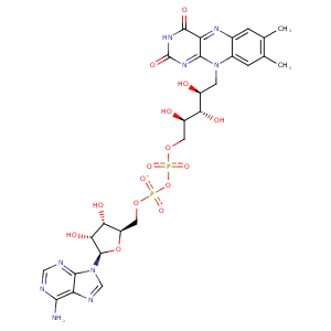
| HET Code: | FDA |
|---|---|
| Formula: | C27H33N9O15P2 |
| Molecular weight: | 785.550 g/mol |
| DrugBank ID: | - |
| Buried Surface Area: | 70.34 % |
| Polar Surface area: | 381.04 Å2 |
| Number of | |
|---|---|
| H-Bond Acceptors: | 21 |
| H-Bond Donors: | 9 |
| Rings: | 6 |
| Aromatic rings: | 3 |
| Anionic atoms: | 2 |
| Cationic atoms: | 0 |
| Rule of Five Violation: | 3 |
| Rotatable Bonds: | 13 |
| X | Y | Z |
|---|---|---|
| 26.9993 | 106.608 | 220.694 |
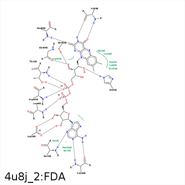


Represent the protein/ligand binding mode, centered on the ligand
Dashed lines represents hydrogen bonds and metal interactions
Green residue labels for amino acids with hydrophobic contacts (green lines) to the ligand
| Ligand | Protein | Interaction | |||
|---|---|---|---|---|---|
| Atom | Atom | Residue | Distance (Å) | Angle (°) | Type |
| C4' | CG | PRO- 17 | 3.78 | 0 | Hydrophobic |
| O1P | N | THR- 18 | 2.99 | 147.08 | H-Bond (Protein Donor) |
| O2P | OG1 | THR- 18 | 2.63 | 153.83 | H-Bond (Protein Donor) |
| O3B | OD2 | ASP- 38 | 2.79 | 178.81 | H-Bond (Ligand Donor) |
| O2B | OD2 | ASP- 38 | 3.4 | 146.64 | H-Bond (Ligand Donor) |
| O2B | OD1 | ASP- 38 | 2.83 | 150.48 | H-Bond (Ligand Donor) |
| N3A | N | SER- 39 | 3.19 | 143.64 | H-Bond (Protein Donor) |
| O2A | N | LEU- 46 | 2.98 | 163.39 | H-Bond (Protein Donor) |
| C8M | CD2 | LEU- 46 | 4.28 | 0 | Hydrophobic |
| C8M | CG1 | VAL- 60 | 4.16 | 0 | Hydrophobic |
| O2' | NE2 | HIS- 63 | 2.94 | 158.53 | H-Bond (Protein Donor) |
| N3 | O | VAL- 64 | 2.85 | 163.52 | H-Bond (Ligand Donor) |
| O4 | N | VAL- 64 | 2.96 | 170.2 | H-Bond (Protein Donor) |
| N6A | O | VAL- 242 | 3.07 | 169.63 | H-Bond (Ligand Donor) |
| N1A | N | VAL- 242 | 2.95 | 171.82 | H-Bond (Protein Donor) |
| C7M | CG2 | THR- 295 | 3.74 | 0 | Hydrophobic |
| C7M | CE1 | TYR- 419 | 3.72 | 0 | Hydrophobic |
| C1' | CD | ARG- 447 | 4.25 | 0 | Hydrophobic |
| C3' | CD | ARG- 447 | 4 | 0 | Hydrophobic |
| C5' | CB | ARG- 447 | 3.83 | 0 | Hydrophobic |
| O2P | N | ARG- 447 | 2.98 | 165.26 | H-Bond (Protein Donor) |
| O3' | O | GLY- 456 | 2.64 | 166.87 | H-Bond (Ligand Donor) |
| N1 | N | GLN- 458 | 3.35 | 135.83 | H-Bond (Protein Donor) |
| O2 | NE2 | GLN- 458 | 3.34 | 139.29 | H-Bond (Protein Donor) |
| O2 | N | GLN- 458 | 2.77 | 159.48 | H-Bond (Protein Donor) |
| C2' | CG | GLN- 458 | 4.02 | 0 | Hydrophobic |
| O3' | OG | SER- 461 | 2.96 | 160.42 | H-Bond (Protein Donor) |
| C5' | CB | SER- 461 | 3.93 | 0 | Hydrophobic |
