sc-PDB
An Annotated Database of Druggable Binding Sites from the Protein DataBank
An Annotated Database of Druggable Binding Sites from the Protein DataBank
1.860 Å
X-ray
2014-01-06
| Name: | R-specific carbonyl reductase |
|---|---|
| ID: | M4VRJ6_CANPA |
| AC: | M4VRJ6 |
| Organism: | Candida parapsilosis |
| Reign: | Eukaryota |
| TaxID: | 5480 |
| EC Number: | / |
| Chain Name: | Percentage of Residues within binding site |
|---|---|
| A | 98 % |
| B | 2 % |
| B-Factor: | 22.144 |
|---|---|
| Number of residues: | 58 |
| Including | |
| Standard Amino Acids: | 53 |
| Non Standard Amino Acids: | 1 |
| Water Molecules: | 4 |
| Cofactors: | |
| Metals: | ZN |
| Ligandability | Volume (Å3) |
|---|---|
| 0.452 | 671.625 |
| % Hydrophobic | % Polar |
|---|---|
| 38.69 | 61.31 |
| According to VolSite | |
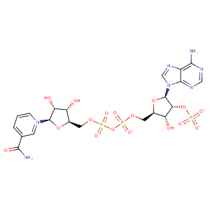
| HET Code: | NDP |
|---|---|
| Formula: | C21H26N7O17P3 |
| Molecular weight: | 741.389 g/mol |
| DrugBank ID: | DB02338 |
| Buried Surface Area: | 72.71 % |
| Polar Surface area: | 404.9 Å2 |
| Number of | |
|---|---|
| H-Bond Acceptors: | 22 |
| H-Bond Donors: | 5 |
| Rings: | 5 |
| Aromatic rings: | 2 |
| Anionic atoms: | 4 |
| Cationic atoms: | 0 |
| Rule of Five Violation: | 2 |
| Rotatable Bonds: | 13 |
| X | Y | Z |
|---|---|---|
| 8.08327 | -9.7906 | -12.3081 |


Represent the protein/ligand binding mode, centered on the ligand
Dashed lines represents hydrogen bonds and metal interactions
Green residue labels for amino acids with hydrophobic contacts (green lines) to the ligand
| Ligand | Protein | Interaction | |||
|---|---|---|---|---|---|
| Atom | Atom | Residue | Distance (Å) | Angle (°) | Type |
| C5N | SG | CYS- 48 | 3.77 | 0 | Hydrophobic |
| O2N | N | TYR- 49 | 2.93 | 165.78 | H-Bond (Protein Donor) |
| C5D | CB | TYR- 49 | 4.49 | 0 | Hydrophobic |
| C3D | CB | TYR- 49 | 4.16 | 0 | Hydrophobic |
| C2D | CB | SER- 50 | 4.43 | 0 | Hydrophobic |
| O2D | OG | SER- 50 | 2.81 | 158.94 | H-Bond (Ligand Donor) |
| C5N | SG | CYS- 167 | 3.56 | 0 | Hydrophobic |
| C4N | CG2 | THR- 171 | 3.6 | 0 | Hydrophobic |
| O3B | O | ILE- 194 | 3.49 | 120.78 | H-Bond (Ligand Donor) |
| O2X | N | ILE- 194 | 2.91 | 164.86 | H-Bond (Protein Donor) |
| O2A | N | GLY- 196 | 2.87 | 176.91 | H-Bond (Protein Donor) |
| O1N | N | LEU- 197 | 2.93 | 173.36 | H-Bond (Protein Donor) |
| C5D | CD1 | LEU- 197 | 4.46 | 0 | Hydrophobic |
| C5N | CD1 | LEU- 197 | 4.03 | 0 | Hydrophobic |
| C4N | CD2 | LEU- 197 | 4.44 | 0 | Hydrophobic |
| O3X | OG | SER- 216 | 3.3 | 138.67 | H-Bond (Protein Donor) |
| O3X | NE | ARG- 217 | 3.01 | 147.67 | H-Bond (Protein Donor) |
| O3X | N | ARG- 217 | 3.25 | 178.65 | H-Bond (Protein Donor) |
| O3X | CZ | ARG- 217 | 3.85 | 0 | Ionic (Protein Cationic) |
| O1X | NZ | LYS- 221 | 2.79 | 153.32 | H-Bond (Protein Donor) |
| O1X | NZ | LYS- 221 | 2.79 | 0 | Ionic (Protein Cationic) |
| O2X | NZ | LYS- 221 | 3.9 | 0 | Ionic (Protein Cationic) |
| C1B | CB | ALA- 257 | 4.33 | 0 | Hydrophobic |
| C5B | CB | SER- 258 | 3.74 | 0 | Hydrophobic |
| C3D | CB | SER- 258 | 4.12 | 0 | Hydrophobic |
| O4B | N | SER- 258 | 3.12 | 157.55 | H-Bond (Protein Donor) |
| N1A | N | ASP- 261 | 2.92 | 130.58 | H-Bond (Protein Donor) |
| N7N | O | VAL- 281 | 3 | 175.97 | H-Bond (Ligand Donor) |
| O3D | O | LEU- 283 | 3.17 | 120.2 | H-Bond (Ligand Donor) |
| O3D | N | LEU- 283 | 3.07 | 158.41 | H-Bond (Protein Donor) |
| C3N | CD1 | LEU- 283 | 4.14 | 0 | Hydrophobic |
| N7N | O | SER- 307 | 3 | 155.98 | H-Bond (Ligand Donor) |
| O7N | N | LEU- 309 | 3 | 155.25 | H-Bond (Protein Donor) |
| C4N | CD2 | LEU- 309 | 4.08 | 0 | Hydrophobic |
| O2N | NH2 | ARG- 356 | 3.35 | 131.32 | H-Bond (Protein Donor) |
| O2N | NH1 | ARG- 356 | 2.74 | 167.52 | H-Bond (Protein Donor) |
| O2N | CZ | ARG- 356 | 3.48 | 0 | Ionic (Protein Cationic) |
| O1N | O | HOH- 512 | 2.85 | 179.95 | H-Bond (Protein Donor) |
