sc-PDB
An Annotated Database of Druggable Binding Sites from the Protein DataBank
An Annotated Database of Druggable Binding Sites from the Protein DataBank
1.200 Å
X-ray
2013-08-09
| Name: | Dihydrofolate reductase |
|---|---|
| ID: | DYR_HUMAN |
| AC: | P00374 |
| Organism: | Homo sapiens |
| Reign: | Eukaryota |
| TaxID: | 9606 |
| EC Number: | 1.5.1.3 |
| Chain Name: | Percentage of Residues within binding site |
|---|---|
| A | 100 % |
| B-Factor: | 22.792 |
|---|---|
| Number of residues: | 44 |
| Including | |
| Standard Amino Acids: | 43 |
| Non Standard Amino Acids: | 0 |
| Water Molecules: | 1 |
| Cofactors: | |
| Metals: | |
| Ligandability | Volume (Å3) |
|---|---|
| 1.173 | 624.375 |
| % Hydrophobic | % Polar |
|---|---|
| 48.65 | 51.35 |
| According to VolSite | |
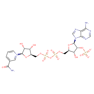
| HET Code: | NDP |
|---|---|
| Formula: | C21H26N7O17P3 |
| Molecular weight: | 741.389 g/mol |
| DrugBank ID: | DB02338 |
| Buried Surface Area: | 65.26 % |
| Polar Surface area: | 404.9 Å2 |
| Number of | |
|---|---|
| H-Bond Acceptors: | 22 |
| H-Bond Donors: | 5 |
| Rings: | 5 |
| Aromatic rings: | 2 |
| Anionic atoms: | 4 |
| Cationic atoms: | 0 |
| Rule of Five Violation: | 2 |
| Rotatable Bonds: | 13 |
| X | Y | Z |
|---|---|---|
| 5.49702 | -3.41833 | -21.6234 |


Represent the protein/ligand binding mode, centered on the ligand
Dashed lines represents hydrogen bonds and metal interactions
Green residue labels for amino acids with hydrophobic contacts (green lines) to the ligand
| Ligand | Protein | Interaction | |||
|---|---|---|---|---|---|
| Atom | Atom | Residue | Distance (Å) | Angle (°) | Type |
| O7N | N | ALA- 9 | 2.98 | 170.49 | H-Bond (Protein Donor) |
| N7N | O | ALA- 9 | 2.84 | 149.98 | H-Bond (Ligand Donor) |
| N7N | O | ILE- 16 | 3.17 | 174.07 | H-Bond (Ligand Donor) |
| C3N | CD2 | LEU- 22 | 3.85 | 0 | Hydrophobic |
| C4B | CB | LYS- 54 | 4.03 | 0 | Hydrophobic |
| C1B | CB | LYS- 54 | 4.29 | 0 | Hydrophobic |
| O4B | N | LYS- 54 | 2.98 | 156.1 | H-Bond (Protein Donor) |
| O2X | NZ | LYS- 54 | 2.52 | 156.56 | H-Bond (Protein Donor) |
| O2X | NZ | LYS- 54 | 2.52 | 0 | Ionic (Protein Cationic) |
| O3X | NZ | LYS- 54 | 3.36 | 0 | Ionic (Protein Cationic) |
| O5B | N | LYS- 55 | 3.22 | 155.57 | H-Bond (Protein Donor) |
| C5B | CG | LYS- 55 | 3.96 | 0 | Hydrophobic |
| O2A | OG1 | THR- 56 | 2.64 | 162.8 | H-Bond (Protein Donor) |
| O2A | N | THR- 56 | 3.02 | 139.32 | H-Bond (Protein Donor) |
| C5N | CG2 | THR- 56 | 4.34 | 0 | Hydrophobic |
| O2X | OG | SER- 76 | 2.52 | 150.41 | H-Bond (Protein Donor) |
| O1X | N | ARG- 77 | 2.81 | 147.56 | H-Bond (Protein Donor) |
| DuAr | CZ | ARG- 77 | 3.84 | 172.08 | Pi/Cation |
| O1A | N | GLY- 117 | 3.1 | 133.1 | H-Bond (Protein Donor) |
| O2A | N | GLY- 117 | 3.06 | 133.78 | H-Bond (Protein Donor) |
| O5D | N | SER- 118 | 2.96 | 141.41 | H-Bond (Protein Donor) |
| O4D | N | SER- 118 | 3.41 | 142.65 | H-Bond (Protein Donor) |
| C4D | CB | SER- 118 | 4.43 | 0 | Hydrophobic |
| O1A | OG | SER- 119 | 2.75 | 135.87 | H-Bond (Protein Donor) |
| O2N | N | SER- 119 | 2.82 | 154.76 | H-Bond (Protein Donor) |
| O2N | OG | SER- 119 | 3.09 | 134.49 | H-Bond (Protein Donor) |
| O3D | O | HOH- 339 | 2.89 | 144.98 | H-Bond (Ligand Donor) |
