sc-PDB
An Annotated Database of Druggable Binding Sites from the Protein DataBank
An Annotated Database of Druggable Binding Sites from the Protein DataBank
2.860 Å
X-ray
2013-08-08
| Name: | UDP-glucuronic acid decarboxylase 1 |
|---|---|
| ID: | UXS1_HUMAN |
| AC: | Q8NBZ7 |
| Organism: | Homo sapiens |
| Reign: | Eukaryota |
| TaxID: | 9606 |
| EC Number: | 4.1.1.35 |
| Chain Name: | Percentage of Residues within binding site |
|---|---|
| B | 98 % |
| C | 2 % |
| B-Factor: | 81.070 |
|---|---|
| Number of residues: | 52 |
| Including | |
| Standard Amino Acids: | 52 |
| Non Standard Amino Acids: | 0 |
| Water Molecules: | 0 |
| Cofactors: | |
| Metals: | |
| Ligandability | Volume (Å3) |
|---|---|
| 1.107 | 1417.500 |
| % Hydrophobic | % Polar |
|---|---|
| 36.67 | 63.33 |
| According to VolSite | |
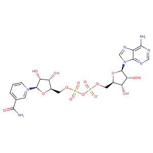
| HET Code: | NAD |
|---|---|
| Formula: | C21H26N7O14P2 |
| Molecular weight: | 662.417 g/mol |
| DrugBank ID: | - |
| Buried Surface Area: | 77.58 % |
| Polar Surface area: | 343.54 Å2 |
| Number of | |
|---|---|
| H-Bond Acceptors: | 18 |
| H-Bond Donors: | 6 |
| Rings: | 5 |
| Aromatic rings: | 3 |
| Anionic atoms: | 2 |
| Cationic atoms: | 1 |
| Rule of Five Violation: | 3 |
| Rotatable Bonds: | 11 |
| X | Y | Z |
|---|---|---|
| -10.3752 | 42.9954 | 344.197 |


Represent the protein/ligand binding mode, centered on the ligand
Dashed lines represents hydrogen bonds and metal interactions
Green residue labels for amino acids with hydrophobic contacts (green lines) to the ligand
| Ligand | Protein | Interaction | |||
|---|---|---|---|---|---|
| Atom | Atom | Residue | Distance (Å) | Angle (°) | Type |
| O1A | N | PHE- 99 | 3.32 | 159.96 | H-Bond (Protein Donor) |
| O1N | N | VAL- 100 | 2.94 | 162.35 | H-Bond (Protein Donor) |
| C5D | CB | VAL- 100 | 4.28 | 0 | Hydrophobic |
| O3B | OD2 | ASP- 119 | 2.51 | 145.2 | H-Bond (Ligand Donor) |
| O3B | OD1 | ASP- 119 | 3.49 | 125.44 | H-Bond (Ligand Donor) |
| O2B | OD1 | ASP- 119 | 2.87 | 136.92 | H-Bond (Ligand Donor) |
| N3A | N | ASN- 120 | 3.36 | 132.73 | H-Bond (Protein Donor) |
| O2A | OG1 | THR- 123 | 2.97 | 169.97 | H-Bond (Protein Donor) |
| O2B | N | THR- 123 | 2.8 | 158.01 | H-Bond (Protein Donor) |
| C2B | CB | THR- 123 | 4.26 | 0 | Hydrophobic |
| O2B | N | GLY- 124 | 2.6 | 141.48 | H-Bond (Protein Donor) |
| N6A | OD1 | ASP- 144 | 2.7 | 139.66 | H-Bond (Ligand Donor) |
| N1A | N | VAL- 145 | 2.8 | 166.03 | H-Bond (Protein Donor) |
| C5D | CB | LEU- 159 | 4.17 | 0 | Hydrophobic |
| C3D | CB | SER- 161 | 4.23 | 0 | Hydrophobic |
| N6A | OG1 | THR- 178 | 2.86 | 133.49 | H-Bond (Ligand Donor) |
| C4D | CB | ALA- 200 | 3.97 | 0 | Hydrophobic |
| C5N | CB | THR- 202 | 3.89 | 0 | Hydrophobic |
| O3D | NZ | LYS- 235 | 2.98 | 129.19 | H-Bond (Protein Donor) |
| C4N | CG2 | ILE- 258 | 4.02 | 0 | Hydrophobic |
| O7N | N | THR- 261 | 3.16 | 155.64 | H-Bond (Protein Donor) |
| O1A | NE2 | HIS- 267 | 2.54 | 142.92 | H-Bond (Protein Donor) |
| O2A | NE2 | HIS- 267 | 3.22 | 133.71 | H-Bond (Protein Donor) |
| O7N | NH1 | ARG- 272 | 2.86 | 150.69 | H-Bond (Protein Donor) |
| O7N | NH2 | ARG- 272 | 3.18 | 135.54 | H-Bond (Protein Donor) |
