sc-PDB
An Annotated Database of Druggable Binding Sites from the Protein DataBank
An Annotated Database of Druggable Binding Sites from the Protein DataBank
2.350 Å
X-ray
2013-07-28
| Name: | Arginine N-methyltransferase, putative |
|---|---|
| ID: | Q57U70_TRYB2 |
| AC: | Q57U70 |
| Organism: | Trypanosoma brucei brucei |
| Reign: | Eukaryota |
| TaxID: | 185431 |
| EC Number: | / |
| Chain Name: | Percentage of Residues within binding site |
|---|---|
| B | 100 % |
| B-Factor: | 22.468 |
|---|---|
| Number of residues: | 35 |
| Including | |
| Standard Amino Acids: | 32 |
| Non Standard Amino Acids: | 1 |
| Water Molecules: | 2 |
| Cofactors: | |
| Metals: | IOD |
| Ligandability | Volume (Å3) |
|---|---|
| 0.875 | 438.750 |
| % Hydrophobic | % Polar |
|---|---|
| 45.38 | 54.62 |
| According to VolSite | |
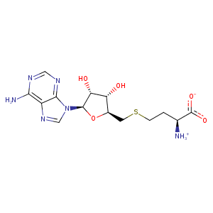
| HET Code: | SAH |
|---|---|
| Formula: | C14H20N6O5S |
| Molecular weight: | 384.411 g/mol |
| DrugBank ID: | DB01752 |
| Buried Surface Area: | 62.89 % |
| Polar Surface area: | 212.38 Å2 |
| Number of | |
|---|---|
| H-Bond Acceptors: | 10 |
| H-Bond Donors: | 4 |
| Rings: | 3 |
| Aromatic rings: | 2 |
| Anionic atoms: | 1 |
| Cationic atoms: | 1 |
| Rule of Five Violation: | 1 |
| Rotatable Bonds: | 7 |
| X | Y | Z |
|---|---|---|
| -26.9755 | 33.6274 | -24.2809 |
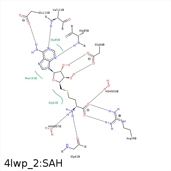


Represent the protein/ligand binding mode, centered on the ligand
Dashed lines represents hydrogen bonds and metal interactions
Green residue labels for amino acids with hydrophobic contacts (green lines) to the ligand
| Ligand | Protein | Interaction | |||
|---|---|---|---|---|---|
| Atom | Atom | Residue | Distance (Å) | Angle (°) | Type |
| SD | CE | MET- 33 | 3.7 | 0 | Hydrophobic |
| CB | CE | MET- 33 | 4 | 0 | Hydrophobic |
| O | NH2 | ARG- 39 | 2.78 | 152.53 | H-Bond (Protein Donor) |
| O | NH1 | ARG- 39 | 3.34 | 128.72 | H-Bond (Protein Donor) |
| OXT | NH2 | ARG- 39 | 3.49 | 127.51 | H-Bond (Protein Donor) |
| OXT | NH1 | ARG- 39 | 2.75 | 162.38 | H-Bond (Protein Donor) |
| O | CZ | ARG- 39 | 3.47 | 0 | Ionic (Protein Cationic) |
| OXT | CZ | ARG- 39 | 3.54 | 0 | Ionic (Protein Cationic) |
| N | O | GLY- 62 | 2.79 | 177.32 | H-Bond (Ligand Donor) |
| O3' | OE1 | GLU- 84 | 3.25 | 127.27 | H-Bond (Ligand Donor) |
| O3' | OE2 | GLU- 84 | 2.61 | 169.04 | H-Bond (Ligand Donor) |
| O2' | OE2 | GLU- 84 | 3.41 | 149.45 | H-Bond (Ligand Donor) |
| N3 | N | ALA- 85 | 3.22 | 141.08 | H-Bond (Protein Donor) |
| N1 | N | VAL- 112 | 2.84 | 162.77 | H-Bond (Protein Donor) |
| N6 | OE1 | GLU- 113 | 2.99 | 160.71 | H-Bond (Ligand Donor) |
| CG | CB | GLU- 142 | 4.49 | 0 | Hydrophobic |
| C1' | SD | MET- 153 | 4.38 | 0 | Hydrophobic |
| C5' | SD | MET- 153 | 3.77 | 0 | Hydrophobic |
| O | O | HOH- 503 | 2.81 | 161.12 | H-Bond (Protein Donor) |
| N | O | HOH- 507 | 3.03 | 160.11 | H-Bond (Ligand Donor) |
