sc-PDB
An Annotated Database of Druggable Binding Sites from the Protein DataBank
An Annotated Database of Druggable Binding Sites from the Protein DataBank
2.000 Å
X-ray
2013-05-22
| Name: | Putative blue-light photoreceptor |
|---|---|
| ID: | A8LP63_DINSH |
| AC: | A8LP63 |
| Organism: | Dinoroseobacter shibae |
| Reign: | Bacteria |
| TaxID: | 398580 |
| EC Number: | / |
| Chain Name: | Percentage of Residues within binding site |
|---|---|
| A | 100 % |
| B-Factor: | 11.097 |
|---|---|
| Number of residues: | 38 |
| Including | |
| Standard Amino Acids: | 35 |
| Non Standard Amino Acids: | 0 |
| Water Molecules: | 3 |
| Cofactors: | |
| Metals: | |
| Ligandability | Volume (Å3) |
|---|---|
| 0.715 | 286.875 |
| % Hydrophobic | % Polar |
|---|---|
| 55.29 | 44.71 |
| According to VolSite | |

| HET Code: | RBF |
|---|---|
| Formula: | C17H20N4O6 |
| Molecular weight: | 376.364 g/mol |
| DrugBank ID: | DB00140 |
| Buried Surface Area: | 80.29 % |
| Polar Surface area: | 155.04 Å2 |
| Number of | |
|---|---|
| H-Bond Acceptors: | 9 |
| H-Bond Donors: | 5 |
| Rings: | 3 |
| Aromatic rings: | 1 |
| Anionic atoms: | 0 |
| Cationic atoms: | 0 |
| Rule of Five Violation: | 0 |
| Rotatable Bonds: | 5 |
| X | Y | Z |
|---|---|---|
| 10.4056 | -2.77967 | -4.76274 |
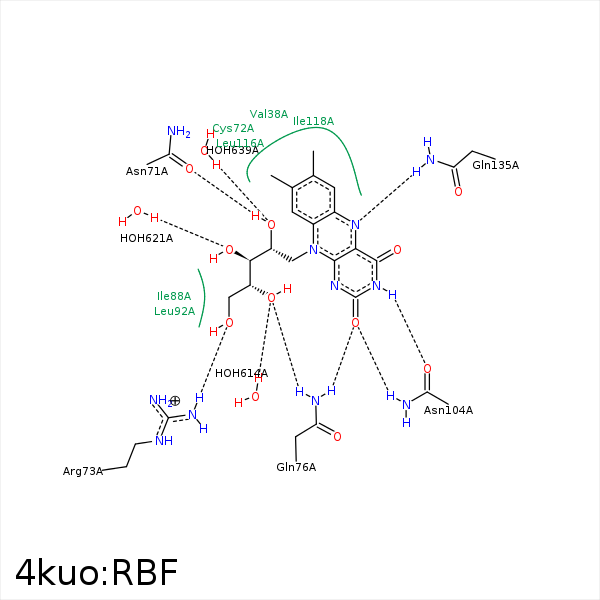


Represent the protein/ligand binding mode, centered on the ligand
Dashed lines represents hydrogen bonds and metal interactions
Green residue labels for amino acids with hydrophobic contacts (green lines) to the ligand
| Ligand | Protein | Interaction | |||
|---|---|---|---|---|---|
| Atom | Atom | Residue | Distance (Å) | Angle (°) | Type |
| C6 | CG2 | VAL- 38 | 3.31 | 0 | Hydrophobic |
| C7M | CB | SER- 40 | 4.13 | 0 | Hydrophobic |
| C8M | CB | SER- 40 | 4.06 | 0 | Hydrophobic |
| O2' | OD1 | ASN- 71 | 2.77 | 168.32 | H-Bond (Ligand Donor) |
| C9A | CB | CYS- 72 | 3.95 | 0 | Hydrophobic |
| C2' | CB | CYS- 72 | 4.48 | 0 | Hydrophobic |
| C2' | CB | ARG- 73 | 4.33 | 0 | Hydrophobic |
| O5' | NH2 | ARG- 73 | 3.09 | 159.18 | H-Bond (Protein Donor) |
| O2 | NE2 | GLN- 76 | 3.18 | 156.05 | H-Bond (Protein Donor) |
| O4' | NE2 | GLN- 76 | 2.8 | 168.43 | H-Bond (Protein Donor) |
| C5' | CG1 | VAL- 85 | 4.09 | 0 | Hydrophobic |
| C1' | CG2 | ILE- 88 | 3.62 | 0 | Hydrophobic |
| C4' | CG2 | ILE- 88 | 3.94 | 0 | Hydrophobic |
| C5' | CB | ARG- 89 | 3.8 | 0 | Hydrophobic |
| C8M | CD2 | LEU- 92 | 3.73 | 0 | Hydrophobic |
| C9 | CD1 | LEU- 92 | 4.48 | 0 | Hydrophobic |
| O2 | ND2 | ASN- 104 | 2.87 | 153.15 | H-Bond (Protein Donor) |
| N3 | OD1 | ASN- 104 | 2.72 | 169.89 | H-Bond (Ligand Donor) |
| C6 | CD1 | LEU- 116 | 4.23 | 0 | Hydrophobic |
| C9A | CD2 | LEU- 116 | 4.26 | 0 | Hydrophobic |
| C9 | CD1 | ILE- 118 | 3.75 | 0 | Hydrophobic |
| C7M | CB | PHE- 131 | 3.94 | 0 | Hydrophobic |
| C8M | CB | PHE- 131 | 3.6 | 0 | Hydrophobic |
| N5 | NE2 | GLN- 135 | 3.02 | 130.86 | H-Bond (Protein Donor) |
| O5' | O | HOH- 614 | 3.44 | 134.43 | H-Bond (Protein Donor) |
| O3' | O | HOH- 621 | 2.74 | 179.95 | H-Bond (Protein Donor) |
| O2' | O | HOH- 639 | 3.06 | 128.42 | H-Bond (Protein Donor) |
