sc-PDB
An Annotated Database of Druggable Binding Sites from the Protein DataBank
An Annotated Database of Druggable Binding Sites from the Protein DataBank
2.360 Å
X-ray
2013-04-24
| Name: | Stimulator of interferon genes protein |
|---|---|
| ID: | STING_MOUSE |
| AC: | Q3TBT3 |
| Organism: | Mus musculus |
| Reign: | Eukaryota |
| TaxID: | 10090 |
| EC Number: | / |
| Chain Name: | Percentage of Residues within binding site |
|---|---|
| A | 49 % |
| B | 51 % |
| B-Factor: | 59.494 |
|---|---|
| Number of residues: | 43 |
| Including | |
| Standard Amino Acids: | 43 |
| Non Standard Amino Acids: | 0 |
| Water Molecules: | 0 |
| Cofactors: | |
| Metals: | |
| Ligandability | Volume (Å3) |
|---|---|
| 0.956 | 1002.375 |
| % Hydrophobic | % Polar |
|---|---|
| 39.39 | 60.61 |
| According to VolSite | |
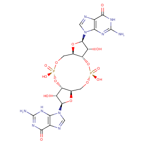
| HET Code: | C2E |
|---|---|
| Formula: | C20H22N10O14P2 |
| Molecular weight: | 688.395 g/mol |
| DrugBank ID: | - |
| Buried Surface Area: | 62.13 % |
| Polar Surface area: | 366.32 Å2 |
| Number of | |
|---|---|
| H-Bond Acceptors: | 20 |
| H-Bond Donors: | 6 |
| Rings: | 7 |
| Aromatic rings: | 2 |
| Anionic atoms: | 2 |
| Cationic atoms: | 0 |
| Rule of Five Violation: | 3 |
| Rotatable Bonds: | 2 |
| X | Y | Z |
|---|---|---|
| -16.7734 | -18.7566 | -7.10489 |
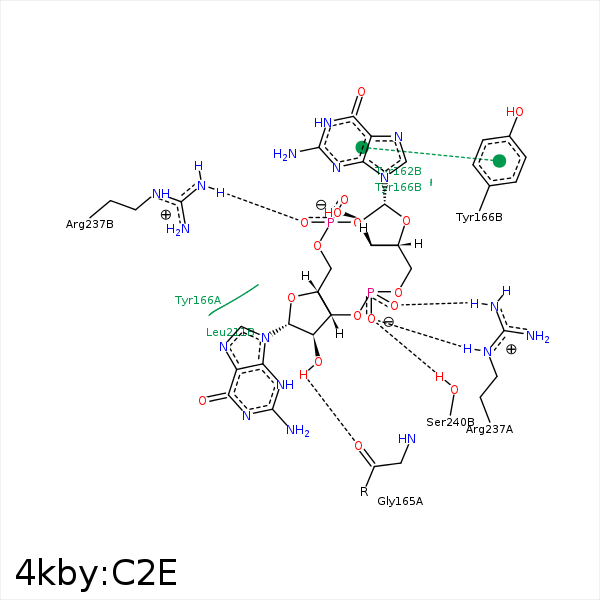


Represent the protein/ligand binding mode, centered on the ligand
Dashed lines represents hydrogen bonds and metal interactions
Green residue labels for amino acids with hydrophobic contacts (green lines) to the ligand
| Ligand | Protein | Interaction | |||
|---|---|---|---|---|---|
| Atom | Atom | Residue | Distance (Å) | Angle (°) | Type |
| O2' | O | GLY- 165 | 3.21 | 156.59 | H-Bond (Ligand Donor) |
| C4A | CZ | TYR- 166 | 4.32 | 0 | Hydrophobic |
| C4' | CE1 | TYR- 166 | 3.5 | 0 | Hydrophobic |
| C1' | CD1 | TYR- 166 | 3.29 | 0 | Hydrophobic |
| C1A | CE1 | TYR- 166 | 3.59 | 0 | Hydrophobic |
| C2' | CD1 | LEU- 211 | 3.92 | 0 | Hydrophobic |
| O2P | NH1 | ARG- 237 | 2.74 | 121.56 | H-Bond (Protein Donor) |
| O21 | NH2 | ARG- 237 | 3.2 | 145.82 | H-Bond (Protein Donor) |
| O11 | NE | ARG- 237 | 3.22 | 144.7 | H-Bond (Protein Donor) |
| O2P | O | VAL- 238 | 2.86 | 164.65 | H-Bond (Ligand Donor) |
| O11 | OG | SER- 240 | 2.87 | 137.61 | H-Bond (Protein Donor) |
| C5A | CB | SER- 240 | 3.82 | 0 | Hydrophobic |
