sc-PDB
An Annotated Database of Druggable Binding Sites from the Protein DataBank
An Annotated Database of Druggable Binding Sites from the Protein DataBank
2.540 Å
X-ray
2013-03-23
| Name: | Voltage-gated potassium channel subunit beta-2 |
|---|---|
| ID: | KCAB2_RAT |
| AC: | P62483 |
| Organism: | Rattus norvegicus |
| Reign: | Eukaryota |
| TaxID: | 10116 |
| EC Number: | / |
| Chain Name: | Percentage of Residues within binding site |
|---|---|
| G | 100 % |
| B-Factor: | 45.126 |
|---|---|
| Number of residues: | 58 |
| Including | |
| Standard Amino Acids: | 56 |
| Non Standard Amino Acids: | 0 |
| Water Molecules: | 2 |
| Cofactors: | |
| Metals: | |
| Ligandability | Volume (Å3) |
|---|---|
| 0.524 | 600.750 |
| % Hydrophobic | % Polar |
|---|---|
| 47.75 | 52.25 |
| According to VolSite | |
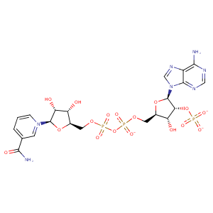
| HET Code: | NAP |
|---|---|
| Formula: | C21H25N7O17P3 |
| Molecular weight: | 740.381 g/mol |
| DrugBank ID: | DB03461 |
| Buried Surface Area: | 82.26 % |
| Polar Surface area: | 405.54 Å2 |
| Number of | |
|---|---|
| H-Bond Acceptors: | 21 |
| H-Bond Donors: | 5 |
| Rings: | 5 |
| Aromatic rings: | 3 |
| Anionic atoms: | 4 |
| Cationic atoms: | 1 |
| Rule of Five Violation: | 2 |
| Rotatable Bonds: | 13 |
| X | Y | Z |
|---|---|---|
| 19.432 | 39.0273 | 219.004 |


Represent the protein/ligand binding mode, centered on the ligand
Dashed lines represents hydrogen bonds and metal interactions
Green residue labels for amino acids with hydrophobic contacts (green lines) to the ligand
| Ligand | Protein | Interaction | |||
|---|---|---|---|---|---|
| Atom | Atom | Residue | Distance (Å) | Angle (°) | Type |
| C5D | CE3 | TRP- 57 | 4.15 | 0 | Hydrophobic |
| C3D | CB | TRP- 57 | 3.54 | 0 | Hydrophobic |
| O3D | N | TRP- 57 | 3.28 | 164.5 | H-Bond (Protein Donor) |
| O2X | NE2 | GLN- 63 | 3.39 | 141.14 | H-Bond (Protein Donor) |
| O2D | OD1 | ASP- 85 | 2.55 | 145.27 | H-Bond (Ligand Donor) |
| C2D | CE2 | TYR- 90 | 3.99 | 0 | Hydrophobic |
| O7N | ND2 | ASN- 158 | 3.28 | 124.23 | H-Bond (Protein Donor) |
| N7N | OG | SER- 188 | 2.77 | 150.03 | H-Bond (Ligand Donor) |
| O7N | NE | ARG- 189 | 2.77 | 145.63 | H-Bond (Protein Donor) |
| O7N | NH2 | ARG- 189 | 2.85 | 139.15 | H-Bond (Protein Donor) |
| N7N | OE1 | GLN- 214 | 2.8 | 149.79 | H-Bond (Ligand Donor) |
| C3N | CB | TRP- 243 | 4.25 | 0 | Hydrophobic |
| C5N | CE3 | TRP- 243 | 3.37 | 0 | Hydrophobic |
| O3 | OG | SER- 244 | 3.49 | 142.4 | H-Bond (Protein Donor) |
| O2N | OG | SER- 244 | 2.5 | 140.16 | H-Bond (Protein Donor) |
| O5D | N | SER- 244 | 3.32 | 130.72 | H-Bond (Protein Donor) |
| O1A | N | LEU- 246 | 2.73 | 146.38 | H-Bond (Protein Donor) |
| O3 | N | CYS- 248 | 3.44 | 134.55 | H-Bond (Protein Donor) |
| N3A | NZ | LYS- 254 | 3.07 | 131.24 | H-Bond (Protein Donor) |
| O1X | NZ | LYS- 254 | 2.68 | 162.16 | H-Bond (Protein Donor) |
| O1X | NZ | LYS- 254 | 2.68 | 0 | Ionic (Protein Cationic) |
| O3B | NH2 | ARG- 264 | 3.38 | 131.44 | H-Bond (Protein Donor) |
| O1N | NH2 | ARG- 264 | 2.84 | 158.53 | H-Bond (Protein Donor) |
| O1N | NH1 | ARG- 264 | 3.28 | 134.56 | H-Bond (Protein Donor) |
| O2N | NH1 | ARG- 264 | 3.13 | 160.8 | H-Bond (Protein Donor) |
| O1N | CZ | ARG- 264 | 3.49 | 0 | Ionic (Protein Cationic) |
| C4D | CB | LEU- 321 | 4.35 | 0 | Hydrophobic |
| C2B | CB | SER- 325 | 4.38 | 0 | Hydrophobic |
| O2B | OG | SER- 325 | 2.62 | 163.67 | H-Bond (Protein Donor) |
| O2B | NE2 | GLN- 329 | 3.05 | 143.15 | H-Bond (Protein Donor) |
| N6A | OE2 | GLU- 332 | 3.26 | 165.56 | H-Bond (Ligand Donor) |
| N7A | ND2 | ASN- 333 | 3.05 | 168.36 | H-Bond (Protein Donor) |
| N6A | OD1 | ASN- 333 | 2.79 | 163.35 | H-Bond (Ligand Donor) |
| O3B | O | HOH- 1136 | 2.67 | 130.88 | H-Bond (Ligand Donor) |
