sc-PDB
An Annotated Database of Druggable Binding Sites from the Protein DataBank
An Annotated Database of Druggable Binding Sites from the Protein DataBank
1.820 Å
X-ray
2013-02-05
| Name: | Kynurenine 3-monooxygenase |
|---|---|
| ID: | KMO_YEAST |
| AC: | P38169 |
| Organism: | Saccharomyces cerevisiae |
| Reign: | Eukaryota |
| TaxID: | 559292 |
| EC Number: | / |
| Chain Name: | Percentage of Residues within binding site |
|---|---|
| B | 100 % |
| B-Factor: | 23.981 |
|---|---|
| Number of residues: | 60 |
| Including | |
| Standard Amino Acids: | 57 |
| Non Standard Amino Acids: | 0 |
| Water Molecules: | 3 |
| Cofactors: | |
| Metals: | |
| Ligandability | Volume (Å3) |
|---|---|
| 0.868 | 1684.125 |
| % Hydrophobic | % Polar |
|---|---|
| 46.09 | 53.91 |
| According to VolSite | |
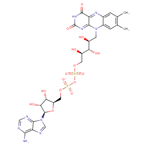
| HET Code: | FAD |
|---|---|
| Formula: | C27H31N9O15P2 |
| Molecular weight: | 783.534 g/mol |
| DrugBank ID: | DB03147 |
| Buried Surface Area: | 66 % |
| Polar Surface area: | 381.7 Å2 |
| Number of | |
|---|---|
| H-Bond Acceptors: | 22 |
| H-Bond Donors: | 7 |
| Rings: | 6 |
| Aromatic rings: | 3 |
| Anionic atoms: | 2 |
| Cationic atoms: | 0 |
| Rule of Five Violation: | 3 |
| Rotatable Bonds: | 13 |
| X | Y | Z |
|---|---|---|
| 41.4144 | 26.8161 | -32.2799 |
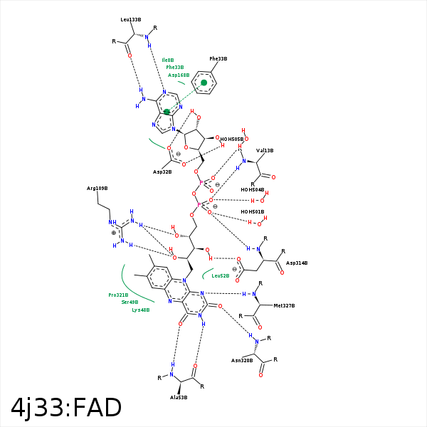


Represent the protein/ligand binding mode, centered on the ligand
Dashed lines represents hydrogen bonds and metal interactions
Green residue labels for amino acids with hydrophobic contacts (green lines) to the ligand
| Ligand | Protein | Interaction | |||
|---|---|---|---|---|---|
| Atom | Atom | Residue | Distance (Å) | Angle (°) | Type |
| C5' | CG2 | VAL- 13 | 4.45 | 0 | Hydrophobic |
| O1P | N | VAL- 13 | 3.08 | 152.62 | H-Bond (Protein Donor) |
| O3B | OD2 | ASP- 32 | 2.58 | 167.6 | H-Bond (Ligand Donor) |
| O2B | OD1 | ASP- 32 | 2.56 | 160.79 | H-Bond (Ligand Donor) |
| O2B | OD2 | ASP- 32 | 3.39 | 129.03 | H-Bond (Ligand Donor) |
| N3A | N | PHE- 33 | 3.29 | 140.17 | H-Bond (Protein Donor) |
| C2B | CE2 | PHE- 33 | 3.76 | 0 | Hydrophobic |
| C8M | CB | LYS- 48 | 3.75 | 0 | Hydrophobic |
| C7M | CB | SER- 49 | 3.91 | 0 | Hydrophobic |
| C2' | CD2 | LEU- 52 | 4.27 | 0 | Hydrophobic |
| N3 | O | ALA- 53 | 3.04 | 156.7 | H-Bond (Ligand Donor) |
| O4 | N | ALA- 53 | 2.8 | 177.22 | H-Bond (Protein Donor) |
| O2' | NH1 | ARG- 109 | 3.02 | 140.51 | H-Bond (Protein Donor) |
| O2' | NH2 | ARG- 109 | 2.87 | 148.95 | H-Bond (Protein Donor) |
| O4' | NH1 | ARG- 109 | 3.15 | 141.95 | H-Bond (Protein Donor) |
| N6A | O | LEU- 133 | 3.18 | 163.57 | H-Bond (Ligand Donor) |
| N1A | N | LEU- 133 | 2.92 | 152.19 | H-Bond (Protein Donor) |
| C1B | CB | ASP- 168 | 4.4 | 0 | Hydrophobic |
| C7M | CZ | TYR- 195 | 3.47 | 0 | Hydrophobic |
| C8M | CE2 | TYR- 195 | 4.01 | 0 | Hydrophobic |
| C8M | CD2 | LEU- 294 | 4.41 | 0 | Hydrophobic |
| O3' | OD1 | ASP- 314 | 2.76 | 162.87 | H-Bond (Ligand Donor) |
| C5' | CB | ASP- 314 | 4.16 | 0 | Hydrophobic |
| O2P | N | ASP- 314 | 2.97 | 164.13 | H-Bond (Protein Donor) |
| C8M | CG | PRO- 321 | 3.96 | 0 | Hydrophobic |
| C8 | CB | PRO- 321 | 3.82 | 0 | Hydrophobic |
| C7M | CZ | PHE- 322 | 3.81 | 0 | Hydrophobic |
| N1 | N | MET- 327 | 3 | 168.64 | H-Bond (Protein Donor) |
| C2' | CB | MET- 327 | 4.12 | 0 | Hydrophobic |
| C4' | CB | MET- 327 | 4.27 | 0 | Hydrophobic |
| O2 | N | ASN- 328 | 2.86 | 165.01 | H-Bond (Protein Donor) |
| O2P | O | HOH- 501 | 2.77 | 179.95 | H-Bond (Protein Donor) |
| O1P | O | HOH- 504 | 2.65 | 179.98 | H-Bond (Protein Donor) |
| O2A | O | HOH- 505 | 2.75 | 179.94 | H-Bond (Protein Donor) |
