sc-PDB
An Annotated Database of Druggable Binding Sites from the Protein DataBank
An Annotated Database of Druggable Binding Sites from the Protein DataBank
1.880 Å
X-ray
2013-01-03
| Name: | Prostaglandin-H2 D-isomerase |
|---|---|
| ID: | PTGDS_HUMAN |
| AC: | P41222 |
| Organism: | Homo sapiens |
| Reign: | Eukaryota |
| TaxID: | 9606 |
| EC Number: | 5.3.99.2 |
| Chain Name: | Percentage of Residues within binding site |
|---|---|
| A | 100 % |
| B-Factor: | 22.579 |
|---|---|
| Number of residues: | 28 |
| Including | |
| Standard Amino Acids: | 27 |
| Non Standard Amino Acids: | 1 |
| Water Molecules: | 0 |
| Cofactors: | |
| Metals: | |
| Ligandability | Volume (Å3) |
|---|---|
| 1.528 | 884.250 |
| % Hydrophobic | % Polar |
|---|---|
| 60.69 | 39.31 |
| According to VolSite | |
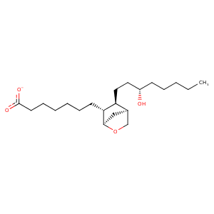
| HET Code: | PWZ |
|---|---|
| Formula: | C21H33O4 |
| Molecular weight: | 349.484 g/mol |
| DrugBank ID: | - |
| Buried Surface Area: | 29.06 % |
| Polar Surface area: | 69.59 Å2 |
| Number of | |
|---|---|
| H-Bond Acceptors: | 4 |
| H-Bond Donors: | 1 |
| Rings: | 2 |
| Aromatic rings: | 0 |
| Anionic atoms: | 1 |
| Cationic atoms: | 0 |
| Rule of Five Violation: | 0 |
| Rotatable Bonds: | 12 |
| X | Y | Z |
|---|---|---|
| 21.469 | 34.9992 | 17.904 |
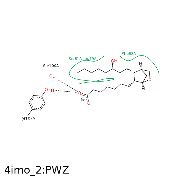


Represent the protein/ligand binding mode, centered on the ligand
Dashed lines represents hydrogen bonds and metal interactions
Green residue labels for amino acids with hydrophobic contacts (green lines) to the ligand
| Ligand | Protein | Interaction | |||
|---|---|---|---|---|---|
| Atom | Atom | Residue | Distance (Å) | Angle (°) | Type |
| C20 | CB | SER- 45 | 3.6 | 0 | Hydrophobic |
| C15 | CB | SER- 81 | 4.27 | 0 | Hydrophobic |
| C17 | CB | SER- 81 | 4.48 | 0 | Hydrophobic |
| C11 | CZ | PHE- 83 | 4.06 | 0 | Hydrophobic |
| C14 | CD1 | PHE- 83 | 4.39 | 0 | Hydrophobic |
| C18 | SD | MET- 94 | 4.45 | 0 | Hydrophobic |
| O2 | OH | TYR- 107 | 2.88 | 163.3 | H-Bond (Protein Donor) |
| O2 | OG | SER- 109 | 2.89 | 141.82 | H-Bond (Protein Donor) |
| C2 | CE2 | TRP- 112 | 4.37 | 0 | Hydrophobic |
| C20 | CE1 | TYR- 149 | 3.92 | 0 | Hydrophobic |
