sc-PDB
An Annotated Database of Druggable Binding Sites from the Protein DataBank
An Annotated Database of Druggable Binding Sites from the Protein DataBank
2.700 Å
X-ray
2012-11-29
| Name: | Diadenosine 5',5'''-P1,P4-tetraphosphate phosphorylase 2 |
|---|---|
| ID: | APA2_YEAST |
| AC: | P22108 |
| Organism: | Saccharomyces cerevisiae |
| Reign: | Eukaryota |
| TaxID: | 559292 |
| EC Number: | / |
| Chain Name: | Percentage of Residues within binding site |
|---|---|
| A | 100 % |
| B-Factor: | 69.737 |
|---|---|
| Number of residues: | 48 |
| Including | |
| Standard Amino Acids: | 46 |
| Non Standard Amino Acids: | 0 |
| Water Molecules: | 2 |
| Cofactors: | |
| Metals: | |
| Ligandability | Volume (Å3) |
|---|---|
| 1.181 | 1447.875 |
| % Hydrophobic | % Polar |
|---|---|
| 45.45 | 54.55 |
| According to VolSite | |
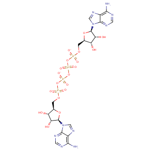
| HET Code: | B4P |
|---|---|
| Formula: | C20H24N10O19P4 |
| Molecular weight: | 832.355 g/mol |
| DrugBank ID: | - |
| Buried Surface Area: | 62.86 % |
| Polar Surface area: | 484.53 Å2 |
| Number of | |
|---|---|
| H-Bond Acceptors: | 27 |
| H-Bond Donors: | 6 |
| Rings: | 6 |
| Aromatic rings: | 4 |
| Anionic atoms: | 4 |
| Cationic atoms: | 0 |
| Rule of Five Violation: | 3 |
| Rotatable Bonds: | 14 |
| X | Y | Z |
|---|---|---|
| 30.9688 | -13.2928 | 21.2313 |
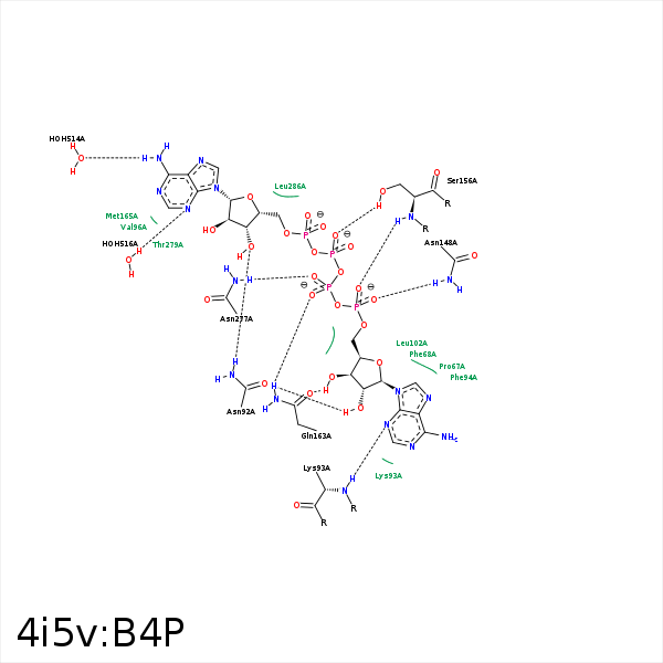


Represent the protein/ligand binding mode, centered on the ligand
Dashed lines represents hydrogen bonds and metal interactions
Green residue labels for amino acids with hydrophobic contacts (green lines) to the ligand
| Ligand | Protein | Interaction | |||
|---|---|---|---|---|---|
| Atom | Atom | Residue | Distance (Å) | Angle (°) | Type |
| O1B | NZ | LYS- 53 | 2.96 | 0 | Ionic (Protein Cationic) |
| C1F | CZ | PHE- 68 | 4.48 | 0 | Hydrophobic |
| C5F | CZ | PHE- 68 | 4.21 | 0 | Hydrophobic |
| O3E | ND2 | ASN- 92 | 2.78 | 126.02 | H-Bond (Protein Donor) |
| O2F | OD1 | ASN- 92 | 2.67 | 154.8 | H-Bond (Ligand Donor) |
| N3B | N | LYS- 93 | 3.25 | 153.68 | H-Bond (Protein Donor) |
| C4E | CG2 | VAL- 96 | 4.21 | 0 | Hydrophobic |
| C1E | CG2 | VAL- 96 | 4.3 | 0 | Hydrophobic |
| C4F | CD2 | LEU- 102 | 4.06 | 0 | Hydrophobic |
| C1F | CD1 | LEU- 102 | 4.43 | 0 | Hydrophobic |
| O1D | ND2 | ASN- 148 | 3.07 | 153.08 | H-Bond (Protein Donor) |
| O1B | OG | SER- 156 | 3.49 | 145.55 | H-Bond (Protein Donor) |
| O2B | OG | SER- 156 | 2.54 | 141.22 | H-Bond (Protein Donor) |
| O2D | N | SER- 156 | 2.98 | 136.12 | H-Bond (Protein Donor) |
| C5F | CB | GLN- 157 | 4.12 | 0 | Hydrophobic |
| O1G | NE2 | GLN- 163 | 3.16 | 149.37 | H-Bond (Protein Donor) |
| O3F | OE1 | GLN- 163 | 2.75 | 162.63 | H-Bond (Ligand Donor) |
| O2G | ND2 | ASN- 277 | 3.19 | 169.1 | H-Bond (Protein Donor) |
| C5E | CD2 | LEU- 286 | 3.93 | 0 | Hydrophobic |
| O1A | NZ | LYS- 288 | 3.5 | 143.94 | H-Bond (Protein Donor) |
| O1A | NZ | LYS- 288 | 3.5 | 0 | Ionic (Protein Cationic) |
| N6A | O | HOH- 514 | 3.2 | 146.62 | H-Bond (Ligand Donor) |
| N3A | O | HOH- 516 | 3.11 | 179.98 | H-Bond (Protein Donor) |
