sc-PDB
An Annotated Database of Druggable Binding Sites from the Protein DataBank
An Annotated Database of Druggable Binding Sites from the Protein DataBank
1.850 Å
X-ray
2012-11-27
| Name: | 1,4-dihydroxy-2-naphthoyl-CoA synthase |
|---|---|
| ID: | MENB_ECOLI |
| AC: | P0ABU0 |
| Organism: | Escherichia coli |
| Reign: | Bacteria |
| TaxID: | 83333 |
| EC Number: | / |
| Chain Name: | Percentage of Residues within binding site |
|---|---|
| B | 2 % |
| C | 16 % |
| E | 82 % |
| B-Factor: | 24.142 |
|---|---|
| Number of residues: | 53 |
| Including | |
| Standard Amino Acids: | 50 |
| Non Standard Amino Acids: | 1 |
| Water Molecules: | 2 |
| Cofactors: | |
| Metals: | CL |
| Ligandability | Volume (Å3) |
|---|---|
| 1.342 | 1073.250 |
| % Hydrophobic | % Polar |
|---|---|
| 57.23 | 42.77 |
| According to VolSite | |
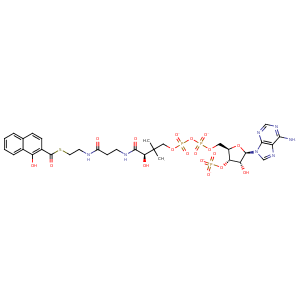
| HET Code: | 1HA |
|---|---|
| Formula: | C32H38N7O18P3S |
| Molecular weight: | 933.667 g/mol |
| DrugBank ID: | - |
| Buried Surface Area: | 58.92 % |
| Polar Surface area: | 449.91 Å2 |
| Number of | |
|---|---|
| H-Bond Acceptors: | 23 |
| H-Bond Donors: | 6 |
| Rings: | 5 |
| Aromatic rings: | 4 |
| Anionic atoms: | 4 |
| Cationic atoms: | 0 |
| Rule of Five Violation: | 3 |
| Rotatable Bonds: | 21 |
| X | Y | Z |
|---|---|---|
| 1.77187 | 30.4078 | -36.6758 |
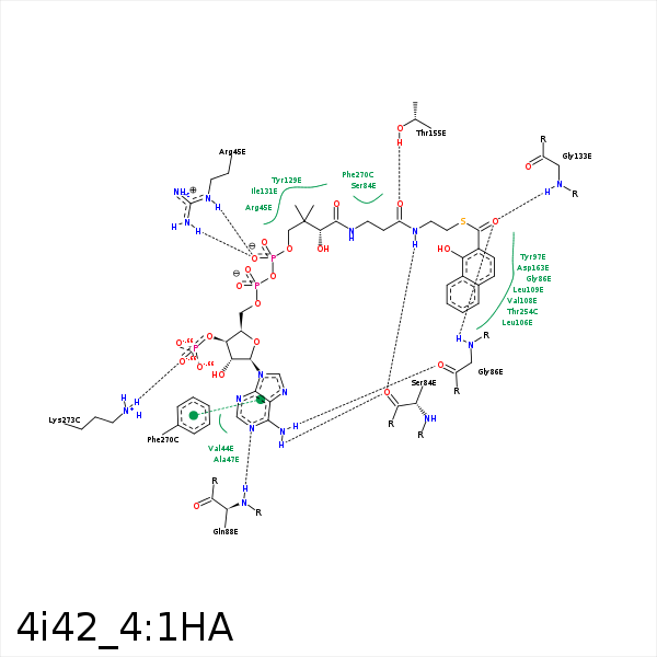


Represent the protein/ligand binding mode, centered on the ligand
Dashed lines represents hydrogen bonds and metal interactions
Green residue labels for amino acids with hydrophobic contacts (green lines) to the ligand
| Ligand | Protein | Interaction | |||
|---|---|---|---|---|---|
| Atom | Atom | Residue | Distance (Å) | Angle (°) | Type |
| CAA | CD1 | ILE- 2 | 4.31 | 0 | Hydrophobic |
| C1' | CB | VAL- 44 | 4.23 | 0 | Hydrophobic |
| CBD | CG | ARG- 45 | 4.13 | 0 | Hydrophobic |
| C5' | CG | ARG- 45 | 4.07 | 0 | Hydrophobic |
| OAP | NH2 | ARG- 45 | 3.22 | 139.68 | H-Bond (Protein Donor) |
| OAP | NE | ARG- 45 | 2.93 | 160.05 | H-Bond (Protein Donor) |
| OBN | NE | ARG- 45 | 3.45 | 121.79 | H-Bond (Protein Donor) |
| OAP | CZ | ARG- 45 | 3.51 | 0 | Ionic (Protein Cationic) |
| NBH | O | SER- 84 | 2.95 | 164.91 | H-Bond (Ligand Donor) |
| N6 | O | SER- 84 | 3.12 | 128.93 | H-Bond (Ligand Donor) |
| CAB | CB | SER- 84 | 3.73 | 0 | Hydrophobic |
| OAF | N | GLY- 86 | 2.91 | 168.43 | H-Bond (Protein Donor) |
| N6 | O | GLY- 86 | 3.02 | 161.33 | H-Bond (Ligand Donor) |
| N1 | N | GLN- 88 | 3.01 | 162.74 | H-Bond (Protein Donor) |
| CAU | CD2 | LEU- 106 | 3.72 | 0 | Hydrophobic |
| CAW | CG1 | VAL- 108 | 3.55 | 0 | Hydrophobic |
| CAQ | CD2 | LEU- 109 | 3.75 | 0 | Hydrophobic |
| CCF | CD1 | TYR- 129 | 4.47 | 0 | Hydrophobic |
| CAA | CE1 | TYR- 129 | 3.87 | 0 | Hydrophobic |
| CAB | CG | TYR- 129 | 4.24 | 0 | Hydrophobic |
| CBD | CE2 | TYR- 129 | 3.84 | 0 | Hydrophobic |
| CBB | CG2 | ILE- 131 | 3.92 | 0 | Hydrophobic |
| CAA | CG1 | ILE- 131 | 4.37 | 0 | Hydrophobic |
| CAB | CG1 | ILE- 131 | 4.12 | 0 | Hydrophobic |
| OAF | N | GLY- 133 | 2.76 | 135.42 | H-Bond (Protein Donor) |
| OAD | OG1 | THR- 155 | 2.62 | 146.05 | H-Bond (Protein Donor) |
| CBB | CG2 | THR- 155 | 4.32 | 0 | Hydrophobic |
| CBA | CG2 | VAL- 159 | 3.95 | 0 | Hydrophobic |
| SBO | CB | SER- 161 | 3.86 | 0 | Hydrophobic |
| CAV | CB | ASP- 163 | 4.13 | 0 | Hydrophobic |
| CAR | CG2 | THR- 254 | 3.5 | 0 | Hydrophobic |
| CBZ | CE1 | PHE- 270 | 4.46 | 0 | Hydrophobic |
| OAN | NZ | LYS- 273 | 2.97 | 149.06 | H-Bond (Protein Donor) |
| OAN | NZ | LYS- 273 | 2.97 | 0 | Ionic (Protein Cationic) |
