sc-PDB
An Annotated Database of Druggable Binding Sites from the Protein DataBank
An Annotated Database of Druggable Binding Sites from the Protein DataBank
2.100 Å
X-ray
2012-09-25
| Name: | Pyridoxine 4-oxidase |
|---|---|
| ID: | Q5NT46_RHILI |
| AC: | Q5NT46 |
| Organism: | Rhizobium loti |
| Reign: | Bacteria |
| TaxID: | 381 |
| EC Number: | / |
| Chain Name: | Percentage of Residues within binding site |
|---|---|
| A | 100 % |
| B-Factor: | 13.865 |
|---|---|
| Number of residues: | 25 |
| Including | |
| Standard Amino Acids: | 24 |
| Non Standard Amino Acids: | 1 |
| Water Molecules: | 0 |
| Cofactors: | FAD |
| Metals: | |
| Ligandability | Volume (Å3) |
|---|---|
| 1.255 | 583.875 |
| % Hydrophobic | % Polar |
|---|---|
| 56.07 | 43.93 |
| According to VolSite | |
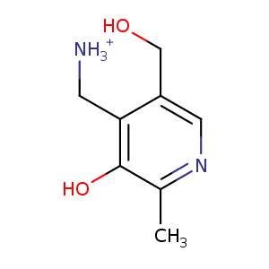
| HET Code: | PXM |
|---|---|
| Formula: | C8H13N2O2 |
| Molecular weight: | 169.201 g/mol |
| DrugBank ID: | DB11673 |
| Buried Surface Area: | 68.48 % |
| Polar Surface area: | 80.99 Å2 |
| Number of | |
|---|---|
| H-Bond Acceptors: | 3 |
| H-Bond Donors: | 3 |
| Rings: | 1 |
| Aromatic rings: | 1 |
| Anionic atoms: | 0 |
| Cationic atoms: | 1 |
| Rule of Five Violation: | 0 |
| Rotatable Bonds: | 2 |
| X | Y | Z |
|---|---|---|
| -30.6993 | 5.04867 | -20.2612 |
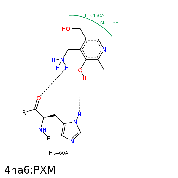


Represent the protein/ligand binding mode, centered on the ligand
Dashed lines represents hydrogen bonds and metal interactions
Green residue labels for amino acids with hydrophobic contacts (green lines) to the ligand
| Ligand | Protein | Interaction | |||
|---|---|---|---|---|---|
| Atom | Atom | Residue | Distance (Å) | Angle (°) | Type |
| C2A | CH2 | TRP- 64 | 3.98 | 0 | Hydrophobic |
| C5 | CB | ALA- 105 | 3.45 | 0 | Hydrophobic |
| C2A | CD1 | LEU- 310 | 4.2 | 0 | Hydrophobic |
| C5A | CB | SER- 330 | 4.04 | 0 | Hydrophobic |
| O3 | ND1 | HIS- 460 | 3.23 | 148.92 | H-Bond (Protein Donor) |
| N4 | O | HIS- 460 | 3.08 | 141.73 | H-Bond (Ligand Donor) |
| N4 | N5 | FAD- 601 | 3.43 | 121.42 | H-Bond (Ligand Donor) |
