sc-PDB
An Annotated Database of Druggable Binding Sites from the Protein DataBank
An Annotated Database of Druggable Binding Sites from the Protein DataBank
1.900 Å
X-ray
2012-07-09
| Name: | Histidinol dehydrogenase |
|---|---|
| ID: | HISX_BRUSU |
| AC: | Q8G2R2 |
| Organism: | Brucella suis biovar 1 |
| Reign: | Bacteria |
| TaxID: | 204722 |
| EC Number: | / |
| Chain Name: | Percentage of Residues within binding site |
|---|---|
| A | 100 % |
| B-Factor: | 37.078 |
|---|---|
| Number of residues: | 26 |
| Including | |
| Standard Amino Acids: | 25 |
| Non Standard Amino Acids: | 1 |
| Water Molecules: | 0 |
| Cofactors: | |
| Metals: | ZN |
| Ligandability | Volume (Å3) |
|---|---|
| 1.151 | 951.750 |
| % Hydrophobic | % Polar |
|---|---|
| 47.16 | 52.84 |
| According to VolSite | |
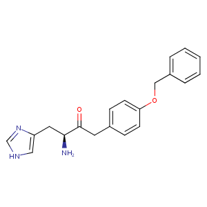
| HET Code: | 0VD |
|---|---|
| Formula: | C20H22N3O2 |
| Molecular weight: | 336.408 g/mol |
| DrugBank ID: | - |
| Buried Surface Area: | 53.41 % |
| Polar Surface area: | 82.62 Å2 |
| Number of | |
|---|---|
| H-Bond Acceptors: | 3 |
| H-Bond Donors: | 2 |
| Rings: | 3 |
| Aromatic rings: | 3 |
| Anionic atoms: | 0 |
| Cationic atoms: | 1 |
| Rule of Five Violation: | 0 |
| Rotatable Bonds: | 8 |
| X | Y | Z |
|---|---|---|
| 57.4061 | 41.8757 | 89.665 |
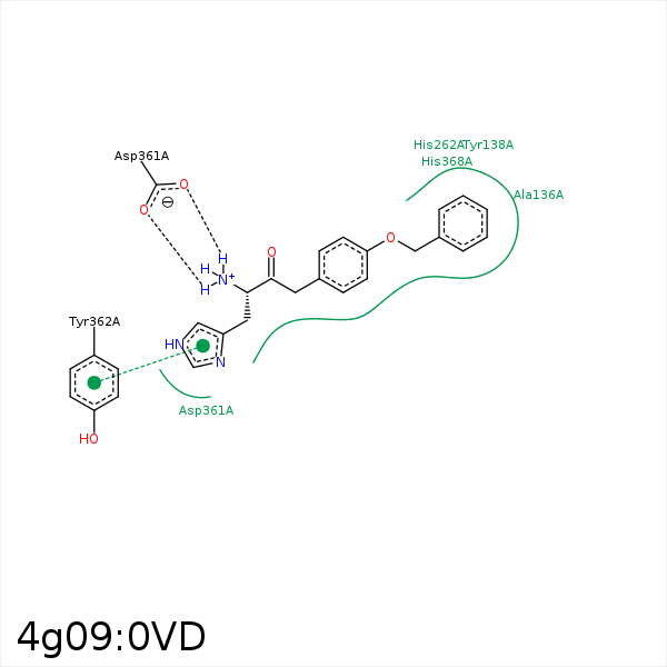


Represent the protein/ligand binding mode, centered on the ligand
Dashed lines represents hydrogen bonds and metal interactions
Green residue labels for amino acids with hydrophobic contacts (green lines) to the ligand
| Ligand | Protein | Interaction | |||
|---|---|---|---|---|---|
| Atom | Atom | Residue | Distance (Å) | Angle (°) | Type |
| C20 | CG | PRO- 132 | 3.48 | 0 | Hydrophobic |
| C19 | CB | ALA- 136 | 3.66 | 0 | Hydrophobic |
| C14 | CE1 | TYR- 138 | 3.45 | 0 | Hydrophobic |
| C10 | CG | PRO- 212 | 4.41 | 0 | Hydrophobic |
| C13 | CB | SER- 237 | 4.29 | 0 | Hydrophobic |
| C7 | CB | SER- 237 | 4.48 | 0 | Hydrophobic |
| C4 | CB | ASP- 361 | 3.87 | 0 | Hydrophobic |
| N3 | OD1 | ASP- 361 | 2.69 | 144.8 | H-Bond (Ligand Donor) |
| C4 | CB | HIS- 368 | 4.03 | 0 | Hydrophobic |
| N2 | ZN | ZN- 501 | 2.19 | 0 | Metal Acceptor |
| DuAr | ZN | ZN- 501 | 3.28 | 103.68 | Pi/Cation |
