sc-PDB
An Annotated Database of Druggable Binding Sites from the Protein DataBank
An Annotated Database of Druggable Binding Sites from the Protein DataBank
2.500 Å
X-ray
2012-04-06
| Name: | Cytochrome P450 2A13 |
|---|---|
| ID: | CP2AD_HUMAN |
| AC: | Q16696 |
| Organism: | Homo sapiens |
| Reign: | Eukaryota |
| TaxID: | 9606 |
| EC Number: | 1.14.14.1 |
| Chain Name: | Percentage of Residues within binding site |
|---|---|
| C | 100 % |
| B-Factor: | 28.965 |
|---|---|
| Number of residues: | 20 |
| Including | |
| Standard Amino Acids: | 19 |
| Non Standard Amino Acids: | 1 |
| Water Molecules: | 0 |
| Cofactors: | |
| Metals: | |
| Ligandability | Volume (Å3) |
|---|---|
| 1.674 | 904.500 |
| % Hydrophobic | % Polar |
|---|---|
| 62.69 | 37.31 |
| According to VolSite | |
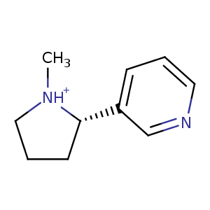
| HET Code: | NCT |
|---|---|
| Formula: | C10H15N2 |
| Molecular weight: | 163.239 g/mol |
| DrugBank ID: | DB00184 |
| Buried Surface Area: | 67.54 % |
| Polar Surface area: | 17.33 Å2 |
| Number of | |
|---|---|
| H-Bond Acceptors: | 1 |
| H-Bond Donors: | 1 |
| Rings: | 2 |
| Aromatic rings: | 1 |
| Anionic atoms: | 0 |
| Cationic atoms: | 1 |
| Rule of Five Violation: | 0 |
| Rotatable Bonds: | 1 |
| X | Y | Z |
|---|---|---|
| 41.1752 | -52.1987 | -38.1073 |
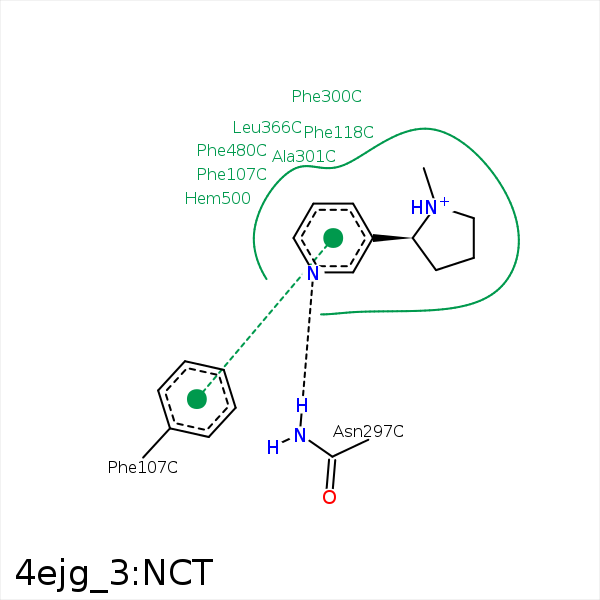


Represent the protein/ligand binding mode, centered on the ligand
Dashed lines represents hydrogen bonds and metal interactions
Green residue labels for amino acids with hydrophobic contacts (green lines) to the ligand
| Ligand | Protein | Interaction | |||
|---|---|---|---|---|---|
| Atom | Atom | Residue | Distance (Å) | Angle (°) | Type |
| C4 | CE1 | PHE- 107 | 3.31 | 0 | Hydrophobic |
| C7 | CZ | PHE- 118 | 4.43 | 0 | Hydrophobic |
| N1 | ND2 | ASN- 297 | 2.96 | 152.61 | H-Bond (Protein Donor) |
| C2 | CB | ALA- 301 | 4.26 | 0 | Hydrophobic |
| C8 | CD2 | LEU- 366 | 3.83 | 0 | Hydrophobic |
| C7 | CD2 | LEU- 370 | 3.81 | 0 | Hydrophobic |
