sc-PDB
An Annotated Database of Druggable Binding Sites from the Protein DataBank
An Annotated Database of Druggable Binding Sites from the Protein DataBank
2.500 Å
X-ray
2012-04-02
| Name: | Mitogen-activated protein kinase 14 |
|---|---|
| ID: | MK14_HUMAN |
| AC: | Q16539 |
| Organism: | Homo sapiens |
| Reign: | Eukaryota |
| TaxID: | 9606 |
| EC Number: | / |
| Chain Name: | Percentage of Residues within binding site |
|---|---|
| A | 100 % |
| B-Factor: | 39.879 |
|---|---|
| Number of residues: | 23 |
| Including | |
| Standard Amino Acids: | 23 |
| Non Standard Amino Acids: | 0 |
| Water Molecules: | 0 |
| Cofactors: | |
| Metals: | |
| Ligandability | Volume (Å3) |
|---|---|
| 1.480 | 347.625 |
| % Hydrophobic | % Polar |
|---|---|
| 72.82 | 27.18 |
| According to VolSite | |
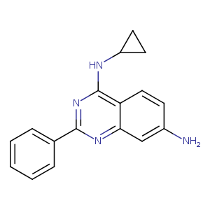
| HET Code: | IRG |
|---|---|
| Formula: | C17H16N4 |
| Molecular weight: | 276.336 g/mol |
| DrugBank ID: | - |
| Buried Surface Area: | 73.68 % |
| Polar Surface area: | 63.83 Å2 |
| Number of | |
|---|---|
| H-Bond Acceptors: | 4 |
| H-Bond Donors: | 2 |
| Rings: | 4 |
| Aromatic rings: | 3 |
| Anionic atoms: | 0 |
| Cationic atoms: | 0 |
| Rule of Five Violation: | 0 |
| Rotatable Bonds: | 3 |
| X | Y | Z |
|---|---|---|
| -23.921 | -15.91 | 9.87405 |
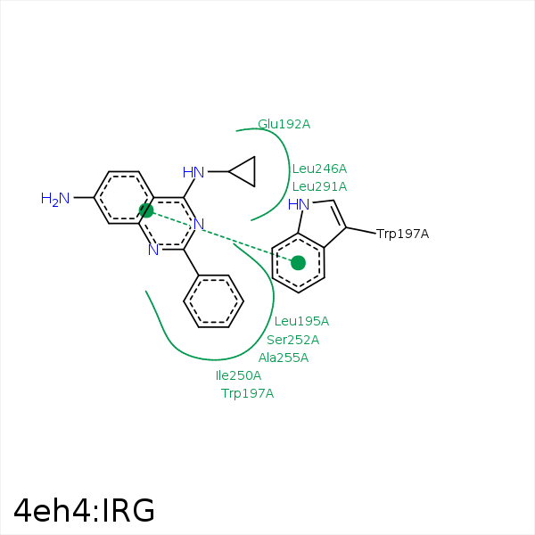


Represent the protein/ligand binding mode, centered on the ligand
Dashed lines represents hydrogen bonds and metal interactions
Green residue labels for amino acids with hydrophobic contacts (green lines) to the ligand
| Ligand | Protein | Interaction | |||
|---|---|---|---|---|---|
| Atom | Atom | Residue | Distance (Å) | Angle (°) | Type |
| CAJ | CB | PRO- 191 | 3.83 | 0 | Hydrophobic |
| C5 | CB | GLU- 192 | 3.91 | 0 | Hydrophobic |
| CAE | CB | LEU- 195 | 3.53 | 0 | Hydrophobic |
| CAK | CD1 | LEU- 195 | 3.34 | 0 | Hydrophobic |
| CAI | CH2 | TRP- 197 | 3.36 | 0 | Hydrophobic |
| CAP | CB | TRP- 197 | 3.7 | 0 | Hydrophobic |
| CAH | CD1 | LEU- 246 | 3.27 | 0 | Hydrophobic |
| CAU | CD2 | LEU- 246 | 3.58 | 0 | Hydrophobic |
| CAI | CB | LYS- 249 | 4.17 | 0 | Hydrophobic |
| CAK | CD1 | ILE- 250 | 4.25 | 0 | Hydrophobic |
| CAF | CG2 | ILE- 250 | 3.98 | 0 | Hydrophobic |
| CAB | CB | SER- 252 | 3.5 | 0 | Hydrophobic |
| CAC | CB | ALA- 255 | 3.74 | 0 | Hydrophobic |
| CAK | CD1 | ILE- 259 | 4.29 | 0 | Hydrophobic |
| CAJ | CG | LEU- 291 | 3.8 | 0 | Hydrophobic |
| CAG | CB | ASP- 292 | 4.44 | 0 | Hydrophobic |
| CAG | CB | SER- 293 | 4.24 | 0 | Hydrophobic |
| NAA | OD1 | ASP- 294 | 3.15 | 126.11 | H-Bond (Ligand Donor) |
| NAA | OD2 | ASP- 294 | 3.25 | 125.47 | H-Bond (Ligand Donor) |
