sc-PDB
An Annotated Database of Druggable Binding Sites from the Protein DataBank
An Annotated Database of Druggable Binding Sites from the Protein DataBank
2.490 Å
X-ray
2012-02-23
| Name: | Cytochrome P450 11B2, mitochondrial |
|---|---|
| ID: | C11B2_HUMAN |
| AC: | P19099 |
| Organism: | Homo sapiens |
| Reign: | Eukaryota |
| TaxID: | 9606 |
| EC Number: | 1.14.15.4 |
| Chain Name: | Percentage of Residues within binding site |
|---|---|
| J | 100 % |
| B-Factor: | 54.396 |
|---|---|
| Number of residues: | 28 |
| Including | |
| Standard Amino Acids: | 27 |
| Non Standard Amino Acids: | 1 |
| Water Molecules: | 0 |
| Cofactors: | |
| Metals: | |
| Ligandability | Volume (Å3) |
|---|---|
| 0.842 | 1728.000 |
| % Hydrophobic | % Polar |
|---|---|
| 55.47 | 44.53 |
| According to VolSite | |
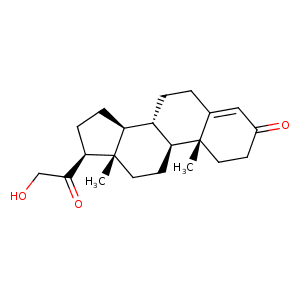
| HET Code: | 1CA |
|---|---|
| Formula: | C21H30O3 |
| Molecular weight: | 330.461 g/mol |
| DrugBank ID: | - |
| Buried Surface Area: | 69.81 % |
| Polar Surface area: | 54.37 Å2 |
| Number of | |
|---|---|
| H-Bond Acceptors: | 3 |
| H-Bond Donors: | 1 |
| Rings: | 4 |
| Aromatic rings: | 0 |
| Anionic atoms: | 0 |
| Cationic atoms: | 0 |
| Rule of Five Violation: | 0 |
| Rotatable Bonds: | 2 |
| X | Y | Z |
|---|---|---|
| 11.3341 | -20.6695 | 58.5201 |
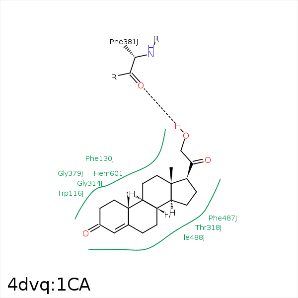


Represent the protein/ligand binding mode, centered on the ligand
Dashed lines represents hydrogen bonds and metal interactions
Green residue labels for amino acids with hydrophobic contacts (green lines) to the ligand
| Ligand | Protein | Interaction | |||
|---|---|---|---|---|---|
| Atom | Atom | Residue | Distance (Å) | Angle (°) | Type |
| C6 | CH2 | TRP- 116 | 4.23 | 0 | Hydrophobic |
| C1 | CG | PHE- 130 | 3.83 | 0 | Hydrophobic |
| C2 | CB | PHE- 130 | 3.97 | 0 | Hydrophobic |
| C12 | CE1 | PHE- 130 | 3.71 | 0 | Hydrophobic |
| C6 | CZ | PHE- 231 | 4.04 | 0 | Hydrophobic |
| C7 | CE1 | PHE- 231 | 3.89 | 0 | Hydrophobic |
| C2 | CB | GLU- 310 | 3.99 | 0 | Hydrophobic |
| C19 | CG2 | THR- 318 | 4.41 | 0 | Hydrophobic |
| C8 | CG2 | THR- 318 | 4.11 | 0 | Hydrophobic |
| O21 | O | PHE- 381 | 2.69 | 156.26 | H-Bond (Ligand Donor) |
| C7 | CE2 | PHE- 487 | 4.09 | 0 | Hydrophobic |
| C15 | CE2 | PHE- 487 | 3.71 | 0 | Hydrophobic |
| C7 | CG2 | ILE- 488 | 4.28 | 0 | Hydrophobic |
| C15 | CG2 | ILE- 488 | 3.71 | 0 | Hydrophobic |
