sc-PDB
An Annotated Database of Druggable Binding Sites from the Protein DataBank
An Annotated Database of Druggable Binding Sites from the Protein DataBank
2.000 Å
X-ray
2013-08-28
| Name: | 3-ketosteroid dehydrogenase |
|---|---|
| ID: | Q9RA02_RHOER |
| AC: | Q9RA02 |
| Organism: | Rhodococcus erythropolis |
| Reign: | Bacteria |
| TaxID: | 1833 |
| EC Number: | / |
| Chain Name: | Percentage of Residues within binding site |
|---|---|
| B | 100 % |
| B-Factor: | 23.180 |
|---|---|
| Number of residues: | 76 |
| Including | |
| Standard Amino Acids: | 70 |
| Non Standard Amino Acids: | 1 |
| Water Molecules: | 5 |
| Cofactors: | |
| Metals: | CL |
| Ligandability | Volume (Å3) |
|---|---|
| 1.289 | 961.875 |
| % Hydrophobic | % Polar |
|---|---|
| 52.63 | 47.37 |
| According to VolSite | |
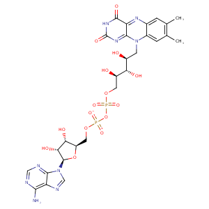
| HET Code: | FAD |
|---|---|
| Formula: | C27H31N9O15P2 |
| Molecular weight: | 783.534 g/mol |
| DrugBank ID: | DB03147 |
| Buried Surface Area: | 76.19 % |
| Polar Surface area: | 381.7 Å2 |
| Number of | |
|---|---|
| H-Bond Acceptors: | 22 |
| H-Bond Donors: | 7 |
| Rings: | 6 |
| Aromatic rings: | 3 |
| Anionic atoms: | 2 |
| Cationic atoms: | 0 |
| Rule of Five Violation: | 3 |
| Rotatable Bonds: | 13 |
| X | Y | Z |
|---|---|---|
| -28.93 | -19.9198 | 60.3205 |


Represent the protein/ligand binding mode, centered on the ligand
Dashed lines represents hydrogen bonds and metal interactions
Green residue labels for amino acids with hydrophobic contacts (green lines) to the ligand
| Ligand | Protein | Interaction | |||
|---|---|---|---|---|---|
| Atom | Atom | Residue | Distance (Å) | Angle (°) | Type |
| O3B | OE1 | GLU- 37 | 2.61 | 151.33 | H-Bond (Ligand Donor) |
| O2B | OE2 | GLU- 37 | 2.81 | 157.09 | H-Bond (Ligand Donor) |
| C1B | CB | LYS- 38 | 4.46 | 0 | Hydrophobic |
| N3A | N | LYS- 38 | 3.2 | 135.03 | H-Bond (Protein Donor) |
| O1A | N | THR- 45 | 2.91 | 154.15 | H-Bond (Protein Donor) |
| O1A | OG1 | THR- 45 | 3.08 | 148.81 | H-Bond (Protein Donor) |
| C4' | CB | THR- 45 | 4.47 | 0 | Hydrophobic |
| C3' | CG2 | THR- 45 | 3.88 | 0 | Hydrophobic |
| C8M | CG2 | THR- 45 | 3.91 | 0 | Hydrophobic |
| O2A | OG | SER- 46 | 2.62 | 155.08 | H-Bond (Protein Donor) |
| O2A | N | SER- 46 | 3.02 | 145.34 | H-Bond (Protein Donor) |
| O4' | OG | SER- 46 | 2.87 | 146.38 | H-Bond (Ligand Donor) |
| C8M | CB | TYR- 48 | 4.18 | 0 | Hydrophobic |
| C6 | CB | SER- 49 | 4.11 | 0 | Hydrophobic |
| C2' | CB | SER- 49 | 4.31 | 0 | Hydrophobic |
| C9A | CB | SER- 49 | 3.62 | 0 | Hydrophobic |
| N5 | N | GLY- 50 | 3.22 | 168.26 | H-Bond (Protein Donor) |
| N3 | O | SER- 52 | 2.7 | 160.48 | H-Bond (Ligand Donor) |
| O4 | N | SER- 52 | 2.95 | 150.96 | H-Bond (Protein Donor) |
| C7M | CD1 | LEU- 153 | 3.88 | 0 | Hydrophobic |
| C8M | CD1 | LEU- 153 | 4.2 | 0 | Hydrophobic |
| N6A | O | LEU- 195 | 3.36 | 164.02 | H-Bond (Ligand Donor) |
| N1A | N | LEU- 195 | 3.07 | 155.1 | H-Bond (Protein Donor) |
| C1B | CB | ALA- 229 | 4.43 | 0 | Hydrophobic |
| C8M | CB | MET- 252 | 3.5 | 0 | Hydrophobic |
| C2B | CB | ALA- 257 | 4.03 | 0 | Hydrophobic |
| O1A | ND2 | ASN- 258 | 3.04 | 161.45 | H-Bond (Protein Donor) |
| N6A | OD2 | ASP- 261 | 2.88 | 140.5 | H-Bond (Ligand Donor) |
| C7M | CD2 | PHE- 294 | 3.76 | 0 | Hydrophobic |
| C7M | CG2 | ILE- 354 | 4.38 | 0 | Hydrophobic |
| C9 | CB | LEU- 447 | 3.8 | 0 | Hydrophobic |
| C9A | CD1 | LEU- 447 | 4.05 | 0 | Hydrophobic |
| C1' | CB | LEU- 447 | 3.85 | 0 | Hydrophobic |
| C8 | CD2 | LEU- 447 | 3.7 | 0 | Hydrophobic |
| O3' | O | LEU- 447 | 2.83 | 167.53 | H-Bond (Ligand Donor) |
| O3' | ND2 | ASN- 477 | 3.27 | 149.66 | H-Bond (Protein Donor) |
| O1P | N | ASN- 477 | 2.91 | 169.17 | H-Bond (Protein Donor) |
| C3' | CB | PRO- 493 | 4.25 | 0 | Hydrophobic |
| O2 | N | LEU- 494 | 2.8 | 153.5 | H-Bond (Protein Donor) |
| C4' | CD2 | LEU- 494 | 4.25 | 0 | Hydrophobic |
| O2P | O | HOH- 2008 | 2.58 | 171.61 | H-Bond (Protein Donor) |
| O3B | O | HOH- 2027 | 3.02 | 156.82 | H-Bond (Protein Donor) |
| N7A | O | HOH- 2029 | 2.71 | 177.96 | H-Bond (Protein Donor) |
| O3P | O | HOH- 2216 | 3.4 | 179.96 | H-Bond (Protein Donor) |
