sc-PDB
An Annotated Database of Druggable Binding Sites from the Protein DataBank
An Annotated Database of Druggable Binding Sites from the Protein DataBank
2.190 Å
X-ray
2013-06-25
| Name: | Ketimine reductase mu-crystallin |
|---|---|
| ID: | CRYM_MOUSE |
| AC: | O54983 |
| Organism: | Mus musculus |
| Reign: | Eukaryota |
| TaxID: | 10090 |
| EC Number: | 1.5.1.25 |
| Chain Name: | Percentage of Residues within binding site |
|---|---|
| B | 100 % |
| B-Factor: | 23.504 |
|---|---|
| Number of residues: | 51 |
| Including | |
| Standard Amino Acids: | 46 |
| Non Standard Amino Acids: | 0 |
| Water Molecules: | 5 |
| Cofactors: | |
| Metals: | |
| Ligandability | Volume (Å3) |
|---|---|
| 1.325 | 1080.000 |
| % Hydrophobic | % Polar |
|---|---|
| 51.25 | 48.75 |
| According to VolSite | |
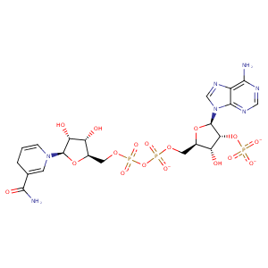
| HET Code: | NDP |
|---|---|
| Formula: | C21H26N7O17P3 |
| Molecular weight: | 741.389 g/mol |
| DrugBank ID: | DB02338 |
| Buried Surface Area: | 62.37 % |
| Polar Surface area: | 404.9 Å2 |
| Number of | |
|---|---|
| H-Bond Acceptors: | 22 |
| H-Bond Donors: | 5 |
| Rings: | 5 |
| Aromatic rings: | 2 |
| Anionic atoms: | 4 |
| Cationic atoms: | 0 |
| Rule of Five Violation: | 2 |
| Rotatable Bonds: | 13 |
| X | Y | Z |
|---|---|---|
| 23.265 | 15.5063 | -7.26404 |


Represent the protein/ligand binding mode, centered on the ligand
Dashed lines represents hydrogen bonds and metal interactions
Green residue labels for amino acids with hydrophobic contacts (green lines) to the ligand
| Ligand | Protein | Interaction | |||
|---|---|---|---|---|---|
| Atom | Atom | Residue | Distance (Å) | Angle (°) | Type |
| O1A | OG | SER- 90 | 2.62 | 169.46 | H-Bond (Protein Donor) |
| O2D | NE2 | HIS- 91 | 3.13 | 148.62 | H-Bond (Ligand Donor) |
| C4N | CG2 | THR- 115 | 3.63 | 0 | Hydrophobic |
| O7N | NH1 | ARG- 118 | 2.65 | 125.08 | H-Bond (Protein Donor) |
| C4N | CG2 | THR- 119 | 3.88 | 0 | Hydrophobic |
| O3B | N | ALA- 143 | 2.92 | 162.78 | H-Bond (Protein Donor) |
| O2A | N | VAL- 145 | 2.95 | 169.32 | H-Bond (Protein Donor) |
| O2N | N | GLN- 146 | 3.02 | 173.61 | H-Bond (Protein Donor) |
| C5D | CG | GLN- 146 | 4.22 | 0 | Hydrophobic |
| O3B | OD1 | ASN- 167 | 2.61 | 131.56 | H-Bond (Ligand Donor) |
| O2B | ND2 | ASN- 167 | 3.46 | 129.7 | H-Bond (Protein Donor) |
| O2X | ND2 | ASN- 167 | 2.52 | 153.59 | H-Bond (Protein Donor) |
| O1X | NH2 | ARG- 168 | 3.09 | 142.52 | H-Bond (Protein Donor) |
| O1X | NE | ARG- 168 | 2.95 | 153.72 | H-Bond (Protein Donor) |
| O3X | NH2 | ARG- 168 | 3.46 | 144.03 | H-Bond (Protein Donor) |
| O1X | CZ | ARG- 168 | 3.45 | 0 | Ionic (Protein Cationic) |
| O1X | OG1 | THR- 169 | 2.78 | 166.56 | H-Bond (Protein Donor) |
| C5D | CB | VAL- 203 | 4.12 | 0 | Hydrophobic |
| C1B | CB | THR- 204 | 4.35 | 0 | Hydrophobic |
| O3D | O | THR- 204 | 3.15 | 164.1 | H-Bond (Ligand Donor) |
| O4B | N | MET- 205 | 3.19 | 127.94 | H-Bond (Protein Donor) |
| C3D | SD | MET- 205 | 3.5 | 0 | Hydrophobic |
| C2D | CE | MET- 205 | 3.96 | 0 | Hydrophobic |
| N7N | O | VAL- 225 | 3.42 | 155.68 | H-Bond (Ligand Donor) |
| N7N | OG | SER- 291 | 3.24 | 151.42 | H-Bond (Ligand Donor) |
| O1N | O | HOH- 2044 | 2.62 | 155.54 | H-Bond (Protein Donor) |
| O2N | O | HOH- 2062 | 2.93 | 179.94 | H-Bond (Protein Donor) |
