sc-PDB
An Annotated Database of Druggable Binding Sites from the Protein DataBank
An Annotated Database of Druggable Binding Sites from the Protein DataBank
2.690 Å
X-ray
2012-05-05
| Name: | Interleukin enhancer-binding factor 2 |
|---|---|
| ID: | ILF2_MOUSE |
| AC: | Q9CXY6 |
| Organism: | Mus musculus |
| Reign: | Eukaryota |
| TaxID: | 10090 |
| EC Number: | / |
| Chain Name: | Percentage of Residues within binding site |
|---|---|
| A | 100 % |
| B-Factor: | 23.059 |
|---|---|
| Number of residues: | 28 |
| Including | |
| Standard Amino Acids: | 27 |
| Non Standard Amino Acids: | 1 |
| Water Molecules: | 0 |
| Cofactors: | |
| Metals: | MG |
| Ligandability | Volume (Å3) |
|---|---|
| 0.637 | 762.750 |
| % Hydrophobic | % Polar |
|---|---|
| 52.65 | 47.35 |
| According to VolSite | |
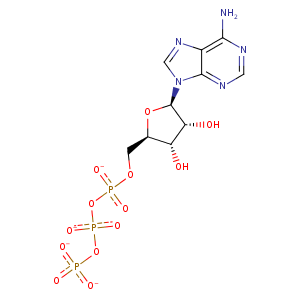
| HET Code: | ATP |
|---|---|
| Formula: | C10H12N5O13P3 |
| Molecular weight: | 503.149 g/mol |
| DrugBank ID: | DB00171 |
| Buried Surface Area: | 43.87 % |
| Polar Surface area: | 319.88 Å2 |
| Number of | |
|---|---|
| H-Bond Acceptors: | 17 |
| H-Bond Donors: | 3 |
| Rings: | 3 |
| Aromatic rings: | 2 |
| Anionic atoms: | 4 |
| Cationic atoms: | 0 |
| Rule of Five Violation: | 2 |
| Rotatable Bonds: | 8 |
| X | Y | Z |
|---|---|---|
| 4.75119 | 0.260677 | -66.0367 |
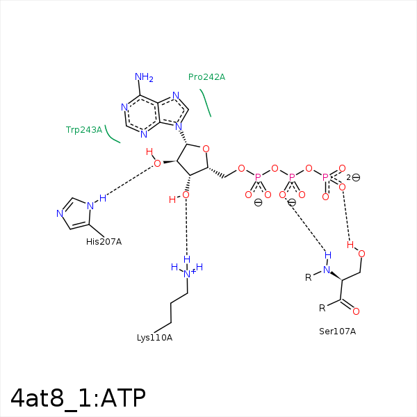


Represent the protein/ligand binding mode, centered on the ligand
Dashed lines represents hydrogen bonds and metal interactions
Green residue labels for amino acids with hydrophobic contacts (green lines) to the ligand
| Ligand | Protein | Interaction | |||
|---|---|---|---|---|---|
| Atom | Atom | Residue | Distance (Å) | Angle (°) | Type |
| O1B | N | SER- 107 | 2.97 | 164.16 | H-Bond (Protein Donor) |
| O2B | NZ | LYS- 110 | 3.93 | 0 | Ionic (Protein Cationic) |
| O3' | NZ | LYS- 110 | 3.05 | 138.68 | H-Bond (Protein Donor) |
| C4' | CB | ALA- 204 | 3.98 | 0 | Hydrophobic |
| O2' | ND1 | HIS- 207 | 2.74 | 162.36 | H-Bond (Protein Donor) |
| O1G | NZ | LYS- 228 | 3.73 | 0 | Ionic (Protein Cationic) |
| O2G | NZ | LYS- 228 | 3.19 | 0 | Ionic (Protein Cationic) |
| O3G | MG | MG- 1361 | 2.21 | 0 | Metal Acceptor |
| O1B | MG | MG- 1361 | 2.58 | 0 | Metal Acceptor |
| O1A | MG | MG- 1361 | 2.64 | 0 | Metal Acceptor |
