sc-PDB
An Annotated Database of Druggable Binding Sites from the Protein DataBank
An Annotated Database of Druggable Binding Sites from the Protein DataBank
1.600 Å
X-ray
2012-05-03
| Name: | Possible succinate dehydrogenase |
|---|---|
| ID: | Q0S4Q9_RHOJR |
| AC: | Q0S4Q9 |
| Organism: | Rhodococcus jostii |
| Reign: | Bacteria |
| TaxID: | 101510 |
| EC Number: | / |
| Chain Name: | Percentage of Residues within binding site |
|---|---|
| A | 100 % |
| B-Factor: | 15.766 |
|---|---|
| Number of residues: | 70 |
| Including | |
| Standard Amino Acids: | 65 |
| Non Standard Amino Acids: | 1 |
| Water Molecules: | 4 |
| Cofactors: | |
| Metals: | CL |
| Ligandability | Volume (Å3) |
|---|---|
| 1.463 | 1022.625 |
| % Hydrophobic | % Polar |
|---|---|
| 56.11 | 43.89 |
| According to VolSite | |
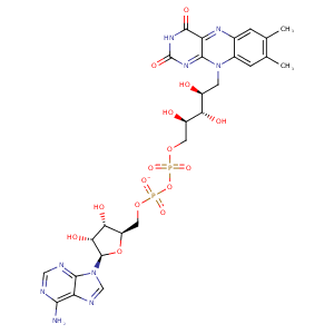
| HET Code: | FAD |
|---|---|
| Formula: | C27H31N9O15P2 |
| Molecular weight: | 783.534 g/mol |
| DrugBank ID: | DB03147 |
| Buried Surface Area: | 73.24 % |
| Polar Surface area: | 381.7 Å2 |
| Number of | |
|---|---|
| H-Bond Acceptors: | 22 |
| H-Bond Donors: | 7 |
| Rings: | 6 |
| Aromatic rings: | 3 |
| Anionic atoms: | 2 |
| Cationic atoms: | 0 |
| Rule of Five Violation: | 3 |
| Rotatable Bonds: | 13 |
| X | Y | Z |
|---|---|---|
| 33.6925 | 30.131 | 40.7055 |


Represent the protein/ligand binding mode, centered on the ligand
Dashed lines represents hydrogen bonds and metal interactions
Green residue labels for amino acids with hydrophobic contacts (green lines) to the ligand
| Ligand | Protein | Interaction | |||
|---|---|---|---|---|---|
| Atom | Atom | Residue | Distance (Å) | Angle (°) | Type |
| O2P | N | ALA- 32 | 3.24 | 168.77 | H-Bond (Protein Donor) |
| O3B | OE1 | GLU- 51 | 2.69 | 169.96 | H-Bond (Ligand Donor) |
| O2B | OE2 | GLU- 51 | 2.71 | 169.91 | H-Bond (Ligand Donor) |
| C1B | CB | ARG- 52 | 4.44 | 0 | Hydrophobic |
| N3A | N | ARG- 52 | 3.27 | 130.25 | H-Bond (Protein Donor) |
| O2A | N | ALA- 59 | 2.83 | 165.69 | H-Bond (Protein Donor) |
| C3' | CB | ALA- 59 | 4.19 | 0 | Hydrophobic |
| O1A | N | THR- 60 | 3.24 | 147.29 | H-Bond (Protein Donor) |
| O1A | OG1 | THR- 60 | 2.62 | 158.16 | H-Bond (Protein Donor) |
| O4' | OG1 | THR- 60 | 2.89 | 143.57 | H-Bond (Ligand Donor) |
| C9A | CB | ALA- 63 | 4.2 | 0 | Hydrophobic |
| C2' | CB | ALA- 63 | 4.45 | 0 | Hydrophobic |
| N5 | N | GLY- 64 | 3.17 | 162.72 | H-Bond (Protein Donor) |
| N3 | O | PHE- 66 | 2.93 | 145.91 | H-Bond (Ligand Donor) |
| O4 | N | PHE- 66 | 2.95 | 141.69 | H-Bond (Protein Donor) |
| C7M | CG2 | THR- 175 | 3.84 | 0 | Hydrophobic |
| N6A | O | VAL- 205 | 3.1 | 158.86 | H-Bond (Ligand Donor) |
| N1A | N | VAL- 205 | 2.9 | 163.34 | H-Bond (Protein Donor) |
| C8M | CB | ALA- 264 | 4.2 | 0 | Hydrophobic |
| C2B | CD1 | ILE- 265 | 3.93 | 0 | Hydrophobic |
| C2B | CB | GLU- 267 | 4.08 | 0 | Hydrophobic |
| O2A | NE2 | HIS- 268 | 2.77 | 171.27 | H-Bond (Protein Donor) |
| C8M | CD2 | PHE- 427 | 4.3 | 0 | Hydrophobic |
| C1' | CZ | PHE- 427 | 3.67 | 0 | Hydrophobic |
| C5' | CG | ARG- 456 | 3.93 | 0 | Hydrophobic |
| O1P | N | ARG- 456 | 2.8 | 168.06 | H-Bond (Protein Donor) |
| O3' | OG | SER- 471 | 2.72 | 155.69 | H-Bond (Protein Donor) |
| C3' | CB | SER- 471 | 4.13 | 0 | Hydrophobic |
| O2 | N | LEU- 472 | 2.76 | 170.94 | H-Bond (Protein Donor) |
| C2' | CD2 | LEU- 472 | 4.15 | 0 | Hydrophobic |
| C4' | CD2 | LEU- 472 | 3.88 | 0 | Hydrophobic |
| O2P | O | HOH- 2039 | 2.81 | 179.96 | H-Bond (Protein Donor) |
| O3B | O | HOH- 2086 | 2.94 | 157.99 | H-Bond (Protein Donor) |
| O1P | O | HOH- 2363 | 2.75 | 159.4 | H-Bond (Protein Donor) |
