sc-PDB
An Annotated Database of Druggable Binding Sites from the Protein DataBank
An Annotated Database of Druggable Binding Sites from the Protein DataBank
2.140 Å
X-ray
2011-07-23
| Name: | Cis-2,3-dihydrobiphenyl-2,3-diol dehydrogenase |
|---|---|
| ID: | BPHB_COMTE |
| AC: | Q46381 |
| Organism: | Comamonas testosteroni |
| Reign: | Bacteria |
| TaxID: | 285 |
| EC Number: | 1.3.1.56 |
| Chain Name: | Percentage of Residues within binding site |
|---|---|
| A | 100 % |
| B-Factor: | 39.856 |
|---|---|
| Number of residues: | 24 |
| Including | |
| Standard Amino Acids: | 23 |
| Non Standard Amino Acids: | 1 |
| Water Molecules: | 0 |
| Cofactors: | NAD |
| Metals: | |
| Ligandability | Volume (Å3) |
|---|---|
| 0.799 | 516.375 |
| % Hydrophobic | % Polar |
|---|---|
| 55.56 | 44.44 |
| According to VolSite | |
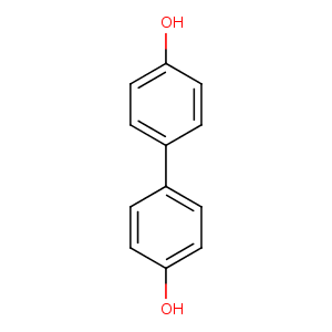
| HET Code: | 4HB |
|---|---|
| Formula: | C12H10O2 |
| Molecular weight: | 186.207 g/mol |
| DrugBank ID: | - |
| Buried Surface Area: | 63.11 % |
| Polar Surface area: | 40.46 Å2 |
| Number of | |
|---|---|
| H-Bond Acceptors: | 2 |
| H-Bond Donors: | 2 |
| Rings: | 2 |
| Aromatic rings: | 2 |
| Anionic atoms: | 0 |
| Cationic atoms: | 0 |
| Rule of Five Violation: | 0 |
| Rotatable Bonds: | 1 |
| X | Y | Z |
|---|---|---|
| 19.9784 | -2.12479 | 40.4125 |


Represent the protein/ligand binding mode, centered on the ligand
Dashed lines represents hydrogen bonds and metal interactions
Green residue labels for amino acids with hydrophobic contacts (green lines) to the ligand
| Ligand | Protein | Interaction | |||
|---|---|---|---|---|---|
| Atom | Atom | Residue | Distance (Å) | Angle (°) | Type |
| CA2 | CZ2 | TRP- 90 | 3.3 | 0 | Hydrophobic |
| OA4 | OG | SER- 142 | 3.11 | 151.18 | H-Bond (Protein Donor) |
| CA2 | CD1 | ILE- 204 | 4.17 | 0 | Hydrophobic |
| CB3 | CD1 | LEU- 209 | 3.21 | 0 | Hydrophobic |
| CB5 | CD2 | LEU- 213 | 3.42 | 0 | Hydrophobic |
| CB6 | CE | MET- 255 | 4.29 | 0 | Hydrophobic |
