sc-PDB
An Annotated Database of Druggable Binding Sites from the Protein DataBank
An Annotated Database of Druggable Binding Sites from the Protein DataBank
2.250 Å
X-ray
2012-12-30
| Min | Mean | Median | Standard Deviation | Max | Count | |
|---|---|---|---|---|---|---|
| pChEMBL: | 7.050 | 7.050 | 7.050 | 0.000 | 7.050 | 1 |
| Name: | 1-deoxy-D-xylulose 5-phosphate reductoisomerase |
|---|---|
| ID: | DXR_MYCTU |
| AC: | P9WNS1 |
| Organism: | Mycobacterium tuberculosis |
| Reign: | Bacteria |
| TaxID: | 83332 |
| EC Number: | / |
| Chain Name: | Percentage of Residues within binding site |
|---|---|
| A | 100 % |
| B-Factor: | 25.103 |
|---|---|
| Number of residues: | 33 |
| Including | |
| Standard Amino Acids: | 32 |
| Non Standard Amino Acids: | 0 |
| Water Molecules: | 1 |
| Cofactors: | |
| Metals: | |
| Ligandability | Volume (Å3) |
|---|---|
| 0.495 | 536.625 |
| % Hydrophobic | % Polar |
|---|---|
| 36.48 | 63.52 |
| According to VolSite | |
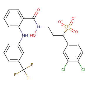
| HET Code: | FM7 |
|---|---|
| Formula: | C23H18Cl2F3N2O5P |
| Molecular weight: | 561.274 g/mol |
| DrugBank ID: | - |
| Buried Surface Area: | 50.1 % |
| Polar Surface area: | 125.57 Å2 |
| Number of | |
|---|---|
| H-Bond Acceptors: | 6 |
| H-Bond Donors: | 2 |
| Rings: | 3 |
| Aromatic rings: | 3 |
| Anionic atoms: | 2 |
| Cationic atoms: | 0 |
| Rule of Five Violation: | 1 |
| Rotatable Bonds: | 9 |
| X | Y | Z |
|---|---|---|
| 20.2842 | 23.4164 | -16.4697 |
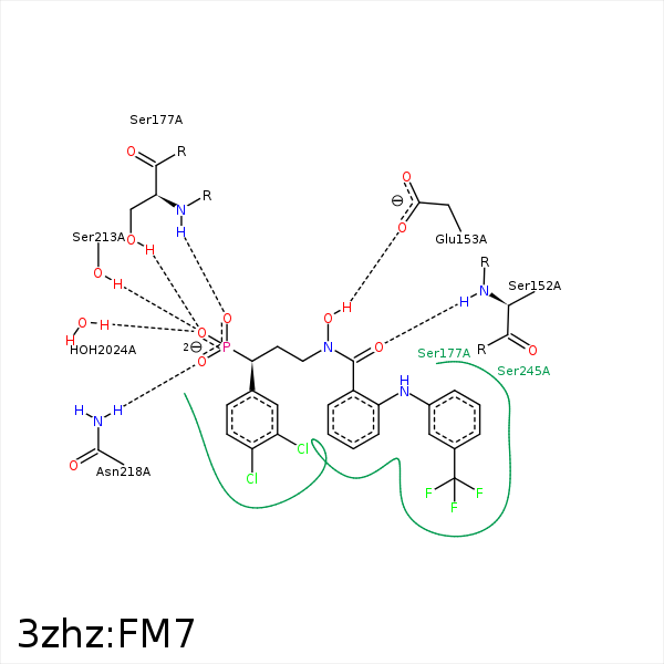


Represent the protein/ligand binding mode, centered on the ligand
Dashed lines represents hydrogen bonds and metal interactions
Green residue labels for amino acids with hydrophobic contacts (green lines) to the ligand
| Ligand | Protein | Interaction | |||
|---|---|---|---|---|---|
| Atom | Atom | Residue | Distance (Å) | Angle (°) | Type |
| F2 | CB | SER- 23 | 3.36 | 0 | Hydrophobic |
| F2 | CD1 | ILE- 24 | 3.54 | 0 | Hydrophobic |
| F3 | CG1 | ILE- 24 | 4.08 | 0 | Hydrophobic |
| O1 | N | SER- 152 | 2.82 | 158.11 | H-Bond (Protein Donor) |
| C17 | CB | SER- 152 | 4.22 | 0 | Hydrophobic |
| O2 | OE1 | GLU- 153 | 2.61 | 162.36 | H-Bond (Ligand Donor) |
| OP3 | OG | SER- 177 | 2.81 | 173.25 | H-Bond (Protein Donor) |
| OP2 | N | SER- 177 | 2.75 | 162.59 | H-Bond (Protein Donor) |
| C11 | CB | SER- 177 | 3.57 | 0 | Hydrophobic |
| F3 | CB | ASN- 209 | 4.42 | 0 | Hydrophobic |
| C21 | CB | ASN- 209 | 4.48 | 0 | Hydrophobic |
| CL1 | CG2 | THR- 210 | 3.9 | 0 | Hydrophobic |
| OP3 | OG | SER- 213 | 2.56 | 157.76 | H-Bond (Protein Donor) |
| C7 | CB | SER- 213 | 3.54 | 0 | Hydrophobic |
| OP1 | ND2 | ASN- 218 | 2.67 | 151.41 | H-Bond (Protein Donor) |
| OP2 | NZ | LYS- 219 | 3.31 | 141.96 | H-Bond (Protein Donor) |
| OP2 | NZ | LYS- 219 | 3.31 | 0 | Ionic (Protein Cationic) |
| OP1 | NZ | LYS- 219 | 3.26 | 0 | Ionic (Protein Cationic) |
| C16 | CB | SER- 245 | 4.38 | 0 | Hydrophobic |
| C15 | CB | PRO- 265 | 4.08 | 0 | Hydrophobic |
| C19 | CG | MET- 267 | 4.5 | 0 | Hydrophobic |
| C18 | SD | MET- 267 | 4.47 | 0 | Hydrophobic |
| C13 | SD | MET- 267 | 3.7 | 0 | Hydrophobic |
| OP3 | O | HOH- 2024 | 2.71 | 179.99 | H-Bond (Protein Donor) |
