sc-PDB
An Annotated Database of Druggable Binding Sites from the Protein DataBank
An Annotated Database of Druggable Binding Sites from the Protein DataBank
2.600 Å
X-ray
2014-10-09
| Name: | Phosphatidylinositol 5-phosphate 4-kinase type-2 beta |
|---|---|
| ID: | PI42B_HUMAN |
| AC: | P78356 |
| Organism: | Homo sapiens |
| Reign: | Eukaryota |
| TaxID: | 9606 |
| EC Number: | 2.7.1.149 |
| Chain Name: | Percentage of Residues within binding site |
|---|---|
| A | 100 % |
| B-Factor: | 68.220 |
|---|---|
| Number of residues: | 20 |
| Including | |
| Standard Amino Acids: | 20 |
| Non Standard Amino Acids: | 0 |
| Water Molecules: | 0 |
| Cofactors: | |
| Metals: | |
| Ligandability | Volume (Å3) |
|---|---|
| 0.525 | 712.125 |
| % Hydrophobic | % Polar |
|---|---|
| 56.87 | 43.13 |
| According to VolSite | |
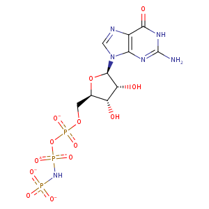
| HET Code: | GNP |
|---|---|
| Formula: | C10H13N6O13P3 |
| Molecular weight: | 518.164 g/mol |
| DrugBank ID: | DB02082 |
| Buried Surface Area: | 41.19 % |
| Polar Surface area: | 338.36 Å2 |
| Number of | |
|---|---|
| H-Bond Acceptors: | 16 |
| H-Bond Donors: | 5 |
| Rings: | 3 |
| Aromatic rings: | 1 |
| Anionic atoms: | 4 |
| Cationic atoms: | 0 |
| Rule of Five Violation: | 2 |
| Rotatable Bonds: | 8 |
| X | Y | Z |
|---|---|---|
| 44.8613 | 61.7976 | -5.16803 |
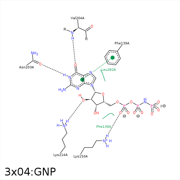


Represent the protein/ligand binding mode, centered on the ligand
Dashed lines represents hydrogen bonds and metal interactions
Green residue labels for amino acids with hydrophobic contacts (green lines) to the ligand
| Ligand | Protein | Interaction | |||
|---|---|---|---|---|---|
| Atom | Atom | Residue | Distance (Å) | Angle (°) | Type |
| C1' | CZ | PHE- 139 | 3.94 | 0 | Hydrophobic |
| O2B | NZ | LYS- 150 | 3.93 | 0 | Ionic (Protein Cationic) |
| O1A | NZ | LYS- 150 | 3.25 | 0 | Ionic (Protein Cationic) |
| O1A | NZ | LYS- 150 | 3.25 | 168.05 | H-Bond (Protein Donor) |
| N1 | OD1 | ASN- 203 | 2.78 | 165.8 | H-Bond (Ligand Donor) |
| O6 | N | VAL- 204 | 3.19 | 155.2 | H-Bond (Protein Donor) |
| O2' | NZ | LYS- 214 | 2.92 | 160.59 | H-Bond (Protein Donor) |
| O3' | OG1 | THR- 237 | 3.49 | 160.99 | H-Bond (Ligand Donor) |
